
Search Count: 20
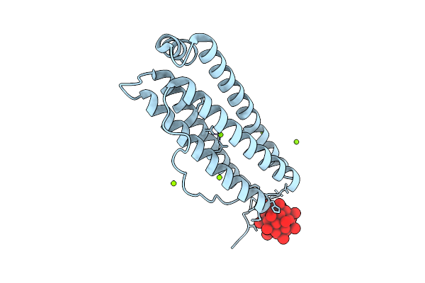 |
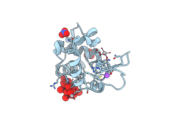
X-Ray Structure Of A Polyoxidovanadate/Human H-Ferritin Adduct Obtained When The Protein Is Treated Overnight With [Vivo(Acac)2]
Organism: Homo sapiens
Method: X-RAY DIFFRACTION Release Date: 2025-12-03 Classification: METAL TRANSPORT Ligands: CL, MG, A1JGJ |
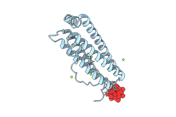 |
X-Ray Structure Of A Polyoxidovanadate/Human H-Ferritin Adduct Obtained When The Protein Is Treated 6 Days With [Vivo(Acac)2]
Organism: Homo sapiens
Method: X-RAY DIFFRACTION Release Date: 2025-12-03 Classification: METAL TRANSPORT Ligands: CL, MG, A1JGJ |
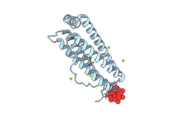 |
X-Ray Structure Of A Polyoxidovanadate/Human H-Ferritin Adduct Obtained When The Protein Is Treated 24 H With [Vivo(Acac)2]
Organism: Homo sapiens
Method: X-RAY DIFFRACTION Release Date: 2025-12-03 Classification: METAL TRANSPORT Ligands: CL, MG, A1JGJ |
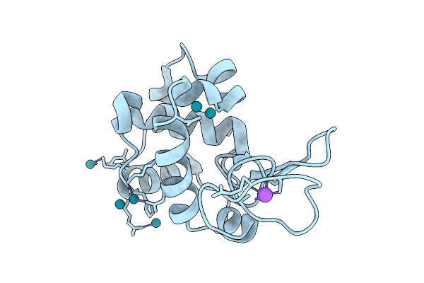 |
X-Ray Structure Of The Adduct Formed Upon Reaction Of Dirhodium-Tetraacetate With Lysozyme At Body Temperature
Organism: Gallus gallus
Method: X-RAY DIFFRACTION Release Date: 2025-08-13 Classification: HYDROLASE Ligands: RH, NA |
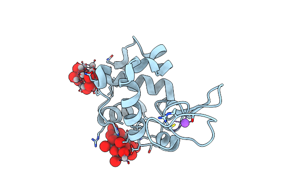 |
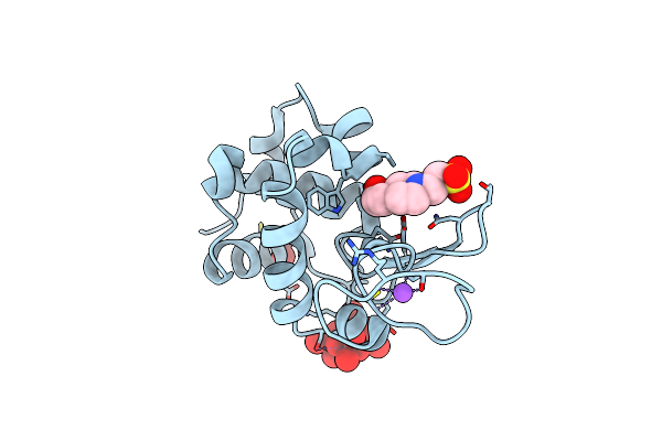
X-Ray Structure Of A Polyoxidovanadate/Lysozyme Adduct Obtained When The Protein Is Treated With [Vivo(Acac)2</Sub>] At 310 K
Organism: Gallus gallus
Method: X-RAY DIFFRACTION Release Date: 2025-05-14 Classification: HYDROLASE Ligands: CL, NA, A1ICR, A1H8D |
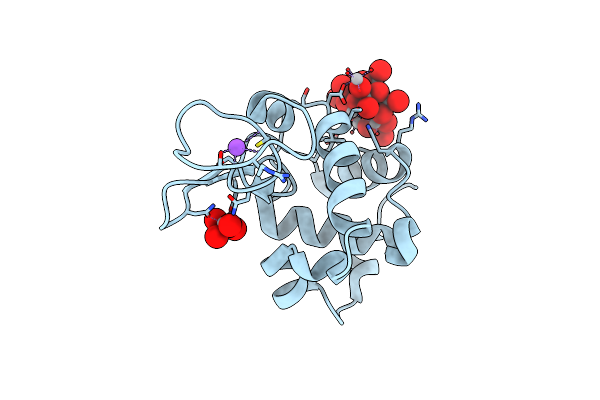 |
X-Ray Structure Of A Polyoxidovanadate/Lysozyme Adduct Obtained When The Protein Is Treated With [Vivo(Acac)2] In 1.1 M Nacl, 0.1 M Sodium Acetate At Ph 4.0 (Structure A)
Organism: Gallus
Method: X-RAY DIFFRACTION Resolution:1.45 Å Release Date: 2024-06-26 Classification: HYDROLASE Ligands: CL, NA, A1ICR, VO4 |
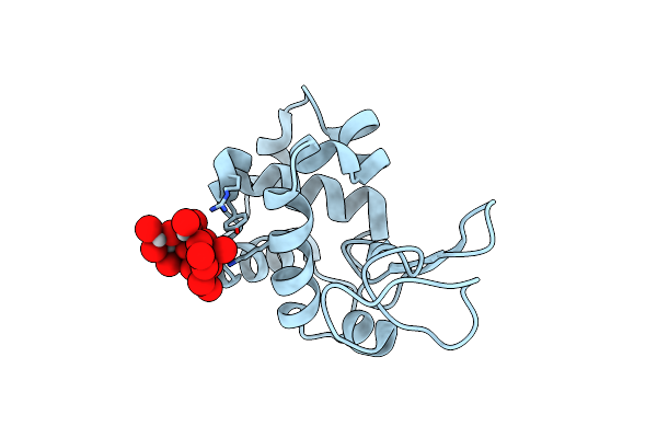 |
X-Ray Structure Of A Polyoxidovanadate/Lysozyme Adduct Obtained When The Protein Is Treated With [Vivo(Acac)2] In 1.1 M Nacl, 0.1 M Sodium Acetate At Ph 4.0 (Structure B)
Organism: Gallus gallus
Method: X-RAY DIFFRACTION Resolution:1.79 Å Release Date: 2024-06-26 Classification: HYDROLASE Ligands: CL, A1H8D |
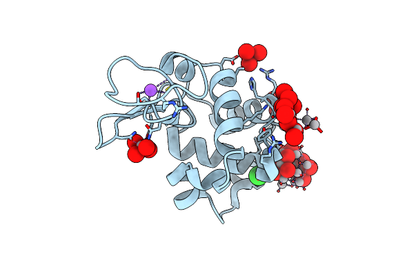 |
X-Ray Structure Of A Polyoxidovanadate/Lysozyme Adduct Obtained When The Protein Is Treated With [Vivo(Acac)2</Sub>] In 1.1 M Nacl, 0.1 M Sodium Acetate At Ph 4.0 (Structure C)
Organism: Gallus gallus
Method: X-RAY DIFFRACTION Resolution:1.17 Å Release Date: 2024-06-26 Classification: HYDROLASE |
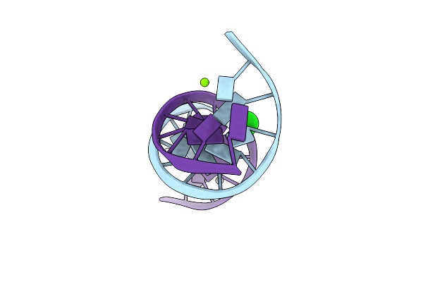 |
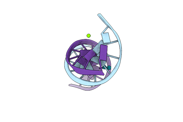
Crystal Structure Of Transplatin/B-Dna Adduct Obtained Upon 7 Days Of Soaking
Organism: Synthetic construct
Method: X-RAY DIFFRACTION Resolution:1.40 Å Release Date: 2024-02-28 Classification: DNA Ligands: MG, PT, NH3, CL |
 |
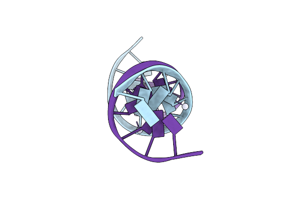
Crystal Structure Of Transplatin/B-Dna Adduct Obtained Upon 48 H Of Soaking
Organism: Dna molecule
Method: X-RAY DIFFRACTION Resolution:1.42 Å Release Date: 2024-02-07 Classification: DNA Ligands: MG, NH3, PT |
 |
Organism: Synthetic construct
Method: X-RAY DIFFRACTION Resolution:2.31 Å Release Date: 2024-01-31 Classification: DNA Ligands: PT |
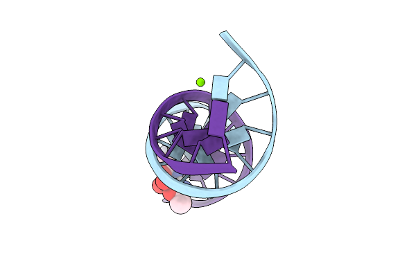 |
Crystal Structure Of Arsenoplatin-1/B-Dna Adduct Obtained Upon 4 H Of Soaking
Organism: Synthetic construct
Method: X-RAY DIFFRACTION Resolution:1.52 Å Release Date: 2024-01-31 Classification: DNA Ligands: PT, MG, A6R |
 |
Crystal Structure Of Arsenoplatin-1/B-Dna Adduct Obtained Upon 48 H Of Soaking
Organism: Synthetic construct
Method: X-RAY DIFFRACTION Resolution:2.51 Å Release Date: 2024-01-31 Classification: DNA Ligands: PT, A6R |
 |
Polyoxidovanadate Interaction With Proteins: Crystal Structure Of Lysozyme Bound To Octadecavanadate Ion (Structure B)
Organism: Gallus gallus
Method: X-RAY DIFFRACTION Resolution:1.80 Å Release Date: 2024-01-10 Classification: HYDROLASE |
 |
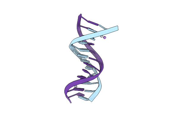
X-Ray Structure Of The Adduct Formed Upon Reaction Of A B-Dna Double Helical Dodecamer With Dirhodium Tetraacetate
Organism: Dna molecule
Method: X-RAY DIFFRACTION Resolution:1.24 Å Release Date: 2023-05-31 Classification: DNA Ligands: MG, RH, CL |
 |
Polyoxidovanadate Interaction With Proteins: Crystal Structure Of Lysozyme Bound To Tetra-Vanadate Ion (Structure 1)
Organism: Gallus gallus
Method: X-RAY DIFFRACTION Resolution:1.18 Å Release Date: 2023-05-24 Classification: HYDROLASE |
 |
X-Ray Structure Of The Adduct Formed Upon Reaction Of The Five-Coordinate Pt(Ii) Complex, 1-Me,Me, With Hewl At Ph 7.5
Organism: Gallus gallus
Method: X-RAY DIFFRACTION Resolution:1.25 Å Release Date: 2023-02-22 Classification: HYDROLASE Ligands: EPE, ACT, R0I, DMS |
 |
X-Ray Structure Of The Adduct Formed Upon Reaction Of The Five-Coordinate Pt(Ii) Complex, 1-Me,Me, With Hewl At Ph 4.0
Organism: Gallus gallus
Method: X-RAY DIFFRACTION Resolution:1.33 Å Release Date: 2023-02-22 Classification: HYDROLASE Ligands: NO3, R0I, PT |
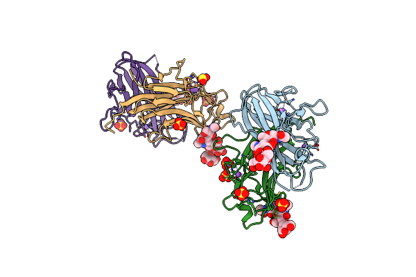 |
Organism: Mus musculus
Method: X-RAY DIFFRACTION Resolution:2.50 Å Release Date: 2018-12-19 Classification: SIGNALING PROTEIN |
 |
Organism: Mus musculus
Method: X-RAY DIFFRACTION Resolution:2.65 Å Release Date: 2018-12-19 Classification: SIGNALING PROTEIN |