
Search Count: 31
 |
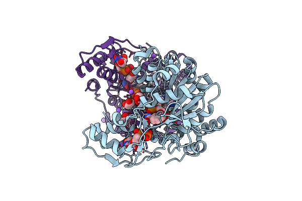
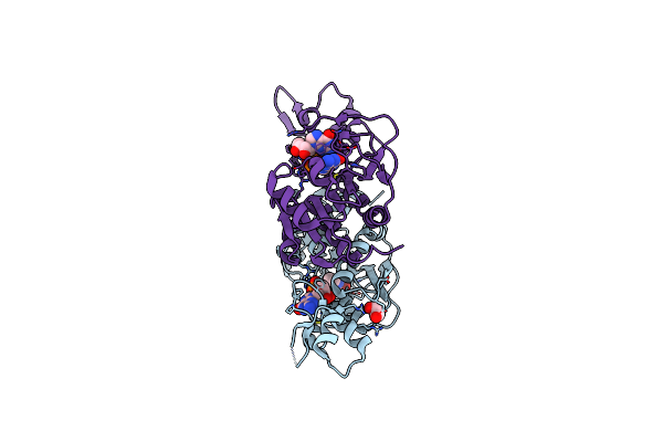
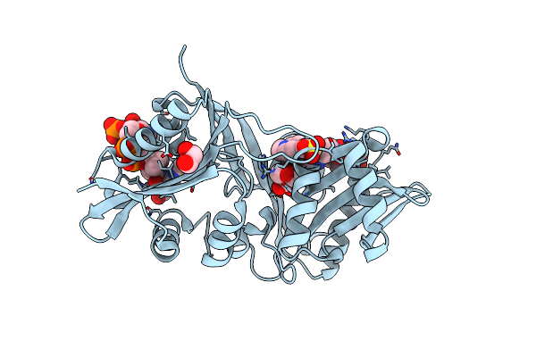
Organism: Mycobacterium tuberculosis (strain atcc 25618 / h37rv)
Method: X-RAY DIFFRACTION Resolution:1.85 Å Release Date: 2018-02-21 Classification: TRANSFERASE Ligands: DAU, MG, TYD, CL, EDO, NA |
 |
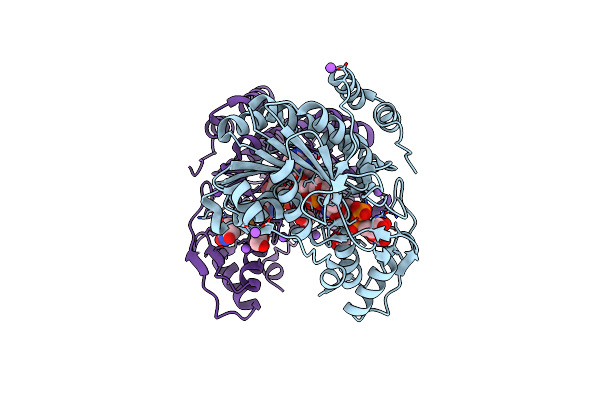
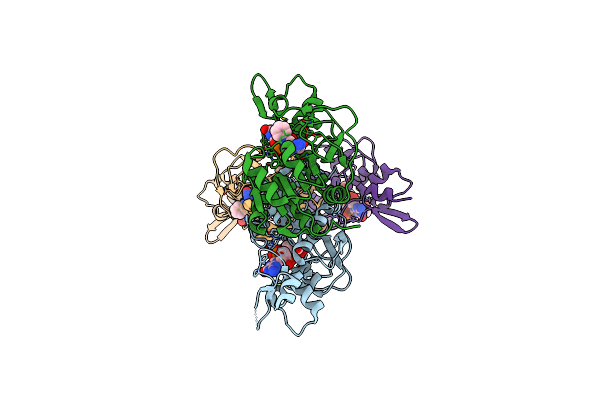
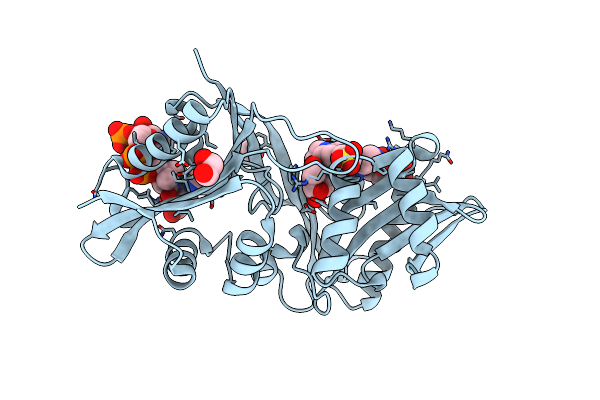
Organism: Mycobacterium tuberculosis (strain atcc 25618 / h37rv)
Method: X-RAY DIFFRACTION Resolution:1.60 Å Release Date: 2018-02-21 Classification: TRANSFERASE Ligands: TTP, MG, TYD, EDO |
 |
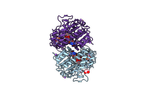
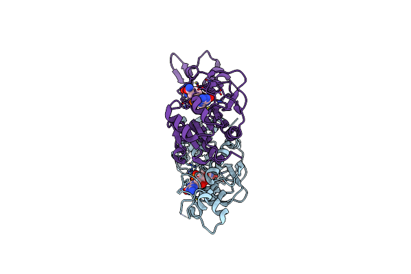
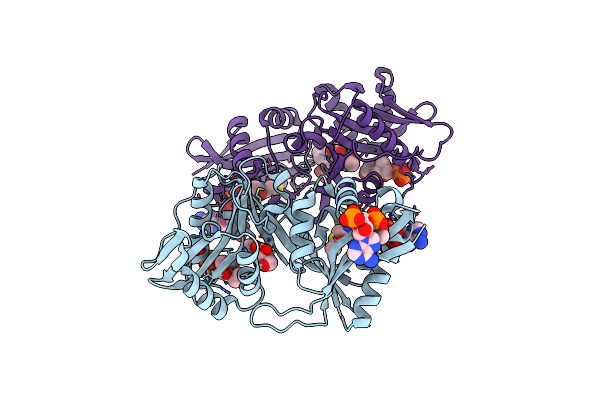
X-Ray Structure Of The Pglf Dehydratase From Campylobacter Jejuni In Complex With Udp And Nad(H)
Organism: Campylobacter jejuni
Method: X-RAY DIFFRACTION Resolution:2.00 Å Release Date: 2017-11-08 Classification: MEMBRANE PROTEIN Ligands: UDP, NAD, EDO, NA |
 |
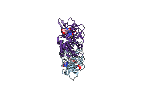
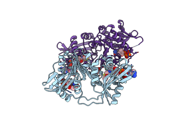
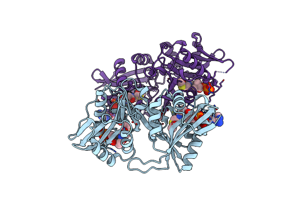
X-Ray Structure Of The Pglf Udp-N-Acetylglucosamine 4,6-Dehydratase From Campylobacterjejuni, D396N/K397A Variant In Complex With Udp-N-Acrtylglucosamine
Organism: Campylobacter jejuni
Method: X-RAY DIFFRACTION Resolution:1.80 Å Release Date: 2017-11-08 Classification: MEMBRANE PROTEIN Ligands: NAD, UD1, EDO, NA |
 |
X-Ray Structure Of The Pglf 4,6-Dehydratase From Campylobacter Jejuni, T595S Variant, In Complex With Udp
Organism: Campylobacter jejuni
Method: X-RAY DIFFRACTION Resolution:1.60 Å Release Date: 2017-11-08 Classification: MEMBRANE PROTEIN Ligands: UDP, NAD, EDO, NA |
 |
X-Ray Structure Of The Pglf 4,6-Dehydratase From Campylobacter Jejuni, Variant T395V, In Complex With Udp
Organism: Campylobacter jejuni
Method: X-RAY DIFFRACTION Resolution:1.60 Å Release Date: 2017-11-08 Classification: MEMBRANE PROTEIN Ligands: UDP, NAD, EDO, NA |
 |
X-Ray Structure Of The Pglf 4,5-Dehydratase From Campylobacter Jejuni, Variant M405Y, In Complex With Udp
Organism: Campylobacter jejuni
Method: X-RAY DIFFRACTION Resolution:1.60 Å Release Date: 2017-11-08 Classification: MEMBRANE PROTEIN Ligands: UDP, NAD, EDO, NA |
 |
Organism: Brucella melitensis biotype 1 (strain 16m / atcc 23456 / nctc 10094)
Method: X-RAY DIFFRACTION Resolution:1.70 Å Release Date: 2017-07-05 Classification: TRANSFERASE Ligands: B62, EDO, CL, GMP |
 |
Crystal Structure Of The Wbkc N-Formyltransferase (C47S Variant) From Brucella Melitensis
Organism: Brucella melitensis biotype 1 (strain 16m / atcc 23456 / nctc 10094)
Method: X-RAY DIFFRACTION Resolution:2.20 Å Release Date: 2017-07-05 Classification: TRANSFERASE Ligands: 1YJ, EDO, GDP, GMP |
 |
Crystal Structure Of The Wbkc N-Formyltransferase (F142A Variant) From Brucella Melitensis
Organism: Brucella melitensis biotype 1 (strain 16m / atcc 23456 / nctc 10094)
Method: X-RAY DIFFRACTION Resolution:2.20 Å Release Date: 2017-07-05 Classification: TRANSFERASE Ligands: FON, GDP, CL |
 |
Crystal Structure Of The Wbkc N-Formyltransferase From Brucella Melitensis In Complex With Gdp-Perosaminea And N-10-Formyltetrahydrofolate
Organism: Brucella melitensis biotype 1 (strain 16m / atcc 23456 / nctc 10094)
Method: X-RAY DIFFRACTION Resolution:2.20 Å Release Date: 2017-07-05 Classification: TRANSFERASE Ligands: 1YA, JB2, GDP |
 |
X-Ray Structure Of A Glucosamine N-Acetyltransferase From Clostridium Acetobutylicum, Apo Form, Ph 5
Organism: Clostridium acetobutylicum (strain atcc 824 / dsm 792 / jcm 1419 / lmg 5710 / vkm b-1787)
Method: X-RAY DIFFRACTION Resolution:2.00 Å Release Date: 2016-07-06 Classification: TRANSFERASE Ligands: ACO, COA, EDO |
 |
X-Ray Structure Of A Glucosamine N-Acetyltransferase From Clostridium Acetobutylicum, Apo Form, Ph 8
Organism: Clostridium acetobutylicum (strain atcc 824 / dsm 792 / jcm 1419 / lmg 5710 / vkm b-1787)
Method: X-RAY DIFFRACTION Resolution:1.90 Å Release Date: 2016-07-06 Classification: TRANSFERASE Ligands: COA, ACO, EDO, PO4 |
 |
X-Ray Structure Of A Glucosamine N-Acetyltransferase From Clostridium Acetobutylicum In Complex With Glucosamine
Organism: Clostridium acetobutylicum (strain atcc 824 / dsm 792 / jcm 1419 / lmg 5710 / vkm b-1787)
Method: X-RAY DIFFRACTION Resolution:1.90 Å Release Date: 2016-07-06 Classification: TRANSFERASE Ligands: ACO, EDO, GCS, EP1, COA |
 |
X-Ray Structure Of A Glucosamine N-Acetyltransferase From Clostridium Acetobutylicum In Complex With N-Acetylglucosamine
Organism: Clostridium acetobutylicum (strain atcc 824 / dsm 792 / jcm 1419 / lmg 5710 / vkm b-1787)
Method: X-RAY DIFFRACTION Resolution:1.49 Å Release Date: 2016-07-06 Classification: TRANSFERASE Ligands: NAG, ACO, COA, EDO, EP1 |
 |
X-Ray Structure Of A Glucosamine N-Acetyltransferase From Clostridium Acetobutylicum, Mutant D287N, In Complex With N-Acetylglucosamine
Organism: Clostridium acetobutylicum (strain atcc 824 / dsm 792 / jcm 1419 / lmg 5710 / vkm b-1787)
Method: X-RAY DIFFRACTION Resolution:1.90 Å Release Date: 2016-07-06 Classification: TRANSFERASE Ligands: NAG, COA, ACO, EDO, CL, TMA |
 |
X-Ray Structure Of A Glucosamine N-Acetyltransferase From Clostridium Acetobutylicum, Mutant Y297F
Organism: Clostridium acetobutylicum (strain atcc 824 / dsm 792 / jcm 1419 / lmg 5710 / vkm b-1787)
Method: X-RAY DIFFRACTION Resolution:1.80 Å Release Date: 2016-07-06 Classification: TRANSFERASE Ligands: ACO, EDO, CL |
 |
X-Ray Structure Of A Glucosamine N-Acetyltransferase From Clostridium Acetobutylicum In Complex With Galactosamine
Organism: Clostridium acetobutylicum (strain atcc 824 / dsm 792 / jcm 1419 / lmg 5710 / vkm b-1787)
Method: X-RAY DIFFRACTION Resolution:1.90 Å Release Date: 2016-07-06 Classification: TRANSFERASE Ligands: COA, X6X, ACO, EDO, EP1 |
 |
X-Ray Structure Of A Glucosamine N-Acetyltransferase From Clostridium Acetobutylicum In Complex With Chitosan
Organism: Clostridium acetobutylicum (strain atcc 824 / dsm 792 / jcm 1419 / lmg 5710 / vkm b-1787)
Method: X-RAY DIFFRACTION Resolution:1.80 Å Release Date: 2016-07-06 Classification: TRANSFERASE Ligands: ACO, COA, EDO, MPO |
 |
Structure Of Pvcb, An Fe, Alpha-Ketoglutarate Dependent Oxygenase From An Isonitrile Synthetic Pathway
Organism: Pseudomonas aeruginosa (pak)
Method: X-RAY DIFFRACTION Resolution:2.05 Å Release Date: 2015-04-29 Classification: OXIDOREDUCTASE Ligands: GOL |