
Search Count: 49
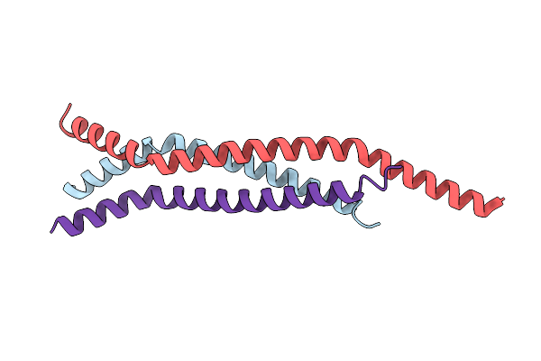 |
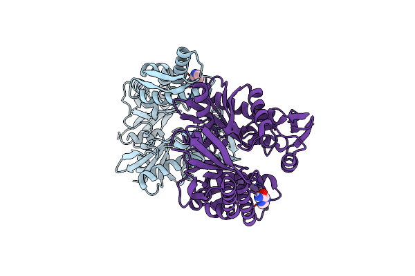
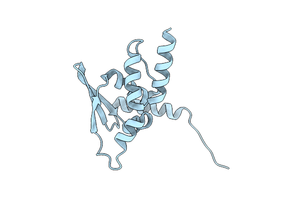
Organism: Lake victoria marburgvirus (strain musoke-80)
Method: X-RAY DIFFRACTION Resolution:2.01 Å Release Date: 2016-11-09 Classification: VIRAL PROTEIN |
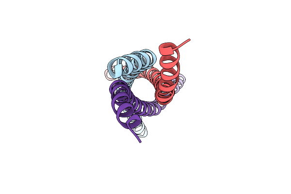 |
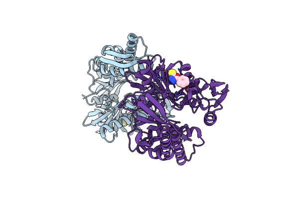
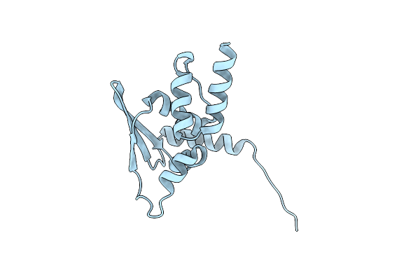
Organism: Lake victoria marburgvirus (strain musoke-80)
Method: X-RAY DIFFRACTION Resolution:2.19 Å Release Date: 2016-11-09 Classification: VIRAL PROTEIN |
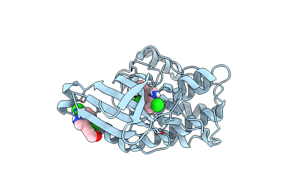 |
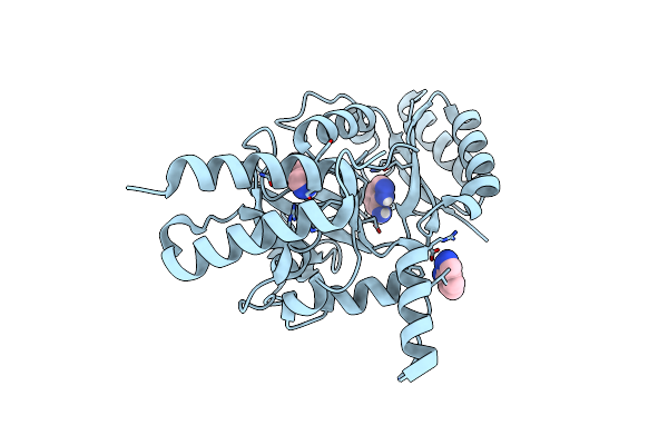
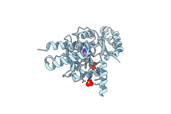
Structure Of Cyclin-Dependent Kinase 2 With Small-Molecule Ligand 3-(4,7-Dichloro-1H-Indol-3-Yl)Prop-2-Yn-1-Ol (At17833) In An Alternate Binding Site.
Organism: Homo sapiens
Method: X-RAY DIFFRACTION Resolution:1.85 Å Release Date: 2015-12-23 Classification: TRANSFERASE Ligands: MFZ |
 |
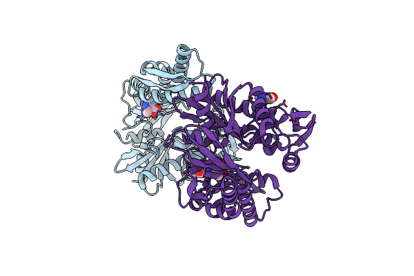
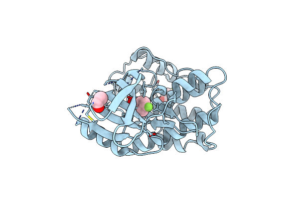
Structure Of Heat Shock-Related 70Kda Protein 2 With Small-Molecule Ligand 1H-1,2,4-Triazol-3-Amine (At485) In An Alternate Binding Site.
Organism: Homo sapiens
Method: X-RAY DIFFRACTION Resolution:1.96 Å Release Date: 2015-12-23 Classification: CHAPERONE Ligands: 3TR |
 |
Structure Of Heat Shock-Related 70Kda Protein 2 With Small-Molecule Ligand 3,5-Dimethyl-1H-Pyrazole-4-Carboxylic Acid (At9084) In An Alternate Binding Site.
Organism: Homo sapiens
Method: X-RAY DIFFRACTION Resolution:1.96 Å Release Date: 2015-12-23 Classification: SIGNALING PROTEIN Ligands: KYD |
 |
Structure Of Bacterial Dna Ligase With Small-Molecule Ligand 1H- Indazol-7-Amine (At4213) In An Alternate Binding Site.
Organism: Staphylococcus aureus
Method: X-RAY DIFFRACTION Resolution:1.83 Å Release Date: 2015-12-23 Classification: LIGASE Ligands: 10L |
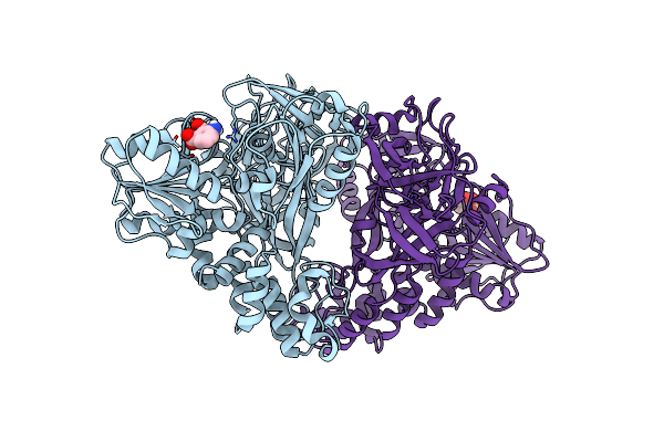 |
Structure Of Hepatitis C Virus (Hcv) Full-Length Ns3 Complex With Small-Molecule Ligand 3-Aminobenzene-1,2-Dicarboxylic Acid (At1246) In An Alternate Binding Site.
Organism: Hepatitis c virus (isolate bk)
Method: X-RAY DIFFRACTION Resolution:2.68 Å Release Date: 2015-12-23 Classification: HYDROLASE Ligands: UP8 |
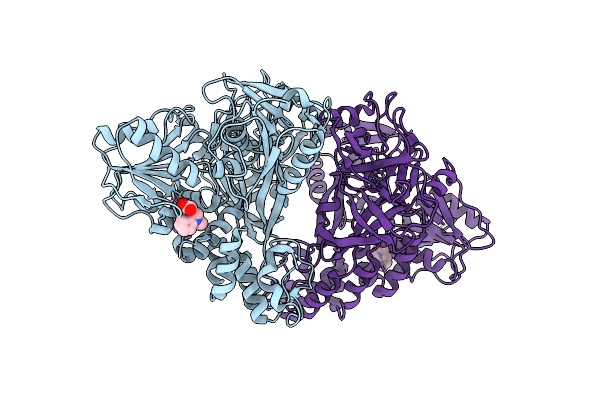 |
Structure Of Hepatitis C Virus (Hcv) Full-Length Ns3 Complex With Small-Molecule Ligand 2-(1-Methyl-1H-Indol-3-Yl)Acetic Acid (At3437) In An Alternate Binding Site.
Organism: Hepatitis c virus (isolate bk)
Method: X-RAY DIFFRACTION Resolution:2.72 Å Release Date: 2015-12-23 Classification: HYDROLASE Ligands: 3VY |
 |
Structure Of Hepatitis C Virus (Hcv) Full-Length Ns3 Complex With Small-Molecule Ligand 5-Bromo-1-Methyl-1H-Indole-2-Carboxylic Acid (At21457) In An Alternate Binding Site.
Organism: Hepatitis c virus (isolate bk)
Method: X-RAY DIFFRACTION Resolution:2.52 Å Release Date: 2015-12-23 Classification: HYDROLASE Ligands: R2N |
 |
Structure Of Heat Shock-Related 70Kda Protein 2 With Small-Molecule Ligand Pyrazine-2-Carboxamide (At513) In An Alternate Binding Site.
Organism: Homo sapiens
Method: X-RAY DIFFRACTION Resolution:1.97 Å Release Date: 2015-12-16 Classification: CHAPERONE Ligands: PZA |
 |
Structure Of Heat Shock-Related 70Kda Protein 2 With Small-Molecule Ligand 5-Phenyl-1,3,4-Oxadiazole-2-Thiol (At809) In An Alternate Binding Site.
Organism: Homo sapiens
Method: X-RAY DIFFRACTION Resolution:1.96 Å Release Date: 2015-12-16 Classification: CHAPERONE Ligands: IWT |
 |
Structure Of Bacterial Dna Ligase With Small-Molecule Ligand Pyrimidin-2-Amine (At371) In An Alternate Binding Site.
Organism: Staphylococcus aureus
Method: X-RAY DIFFRACTION Resolution:2.00 Å Release Date: 2015-12-16 Classification: LIGASE Ligands: SO4, LGA |
 |
Structure Of Cyclin-Dependent Kinase 2 With Small-Molecule Ligand 4- Fluorobenzoic Acid (At222) In An Alternate Binding Site.
Organism: Homo sapiens
Method: X-RAY DIFFRACTION Resolution:2.16 Å Release Date: 2015-12-09 Classification: TRANSFERASE Ligands: 1Y6, ACE |
 |
Organism: Homo sapiens
Method: X-RAY DIFFRACTION Resolution:2.35 Å Release Date: 2014-06-18 Classification: TRANSCRIPTION |
 |
Organism: Homo sapiens
Method: X-RAY DIFFRACTION Resolution:2.80 Å Release Date: 2014-06-18 Classification: TRANSCRIPTION |
 |
Organism: Homo sapiens
Method: X-RAY DIFFRACTION Resolution:2.66 Å Release Date: 2014-06-18 Classification: SIGNALING PROTEIN Ligands: SXJ |
 |
Organism: Homo sapiens
Method: X-RAY DIFFRACTION Resolution:2.30 Å Release Date: 2009-03-10 Classification: CELL CYCLE Ligands: GOL |
 |
Organism: Homo sapiens
Method: X-RAY DIFFRACTION Resolution:2.80 Å Release Date: 2009-03-10 Classification: CELL CYCLE |
 |
Organism: Homo sapiens
Method: X-RAY DIFFRACTION Resolution:2.85 Å Release Date: 2009-03-10 Classification: CELL CYCLE |
 |
Organism: Homo sapiens
Method: X-RAY DIFFRACTION Resolution:2.45 Å Release Date: 2009-03-10 Classification: TRANSFERASE |