
Search Count: 15
 |
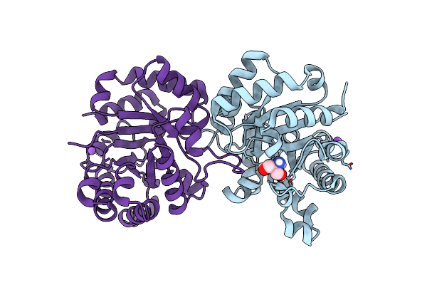
Organism: Rhodonellum psychrophilum
Method: X-RAY DIFFRACTION Resolution:1.50 Å Release Date: 2025-05-14 Classification: ISOMERASE |
 |
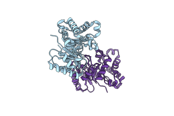
Organism: Rhodococcus sp. jg-3
Method: X-RAY DIFFRACTION Resolution:1.63 Å Release Date: 2025-05-14 Classification: ISOMERASE Ligands: TRS, NA |
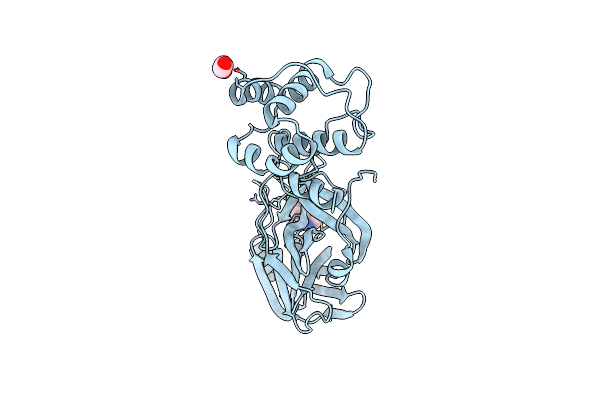 |
Organism: Severe acute respiratory syndrome coronavirus 2
Method: X-RAY DIFFRACTION Resolution:2.70 Å Release Date: 2023-09-27 Classification: VIRAL PROTEIN Ligands: EDO, KM6 |
 |
Crystal Structure Of Human 3-Phosphoglycerate Dehydrogenase In Complex With Gdd-04-35
Organism: Homo sapiens
Method: X-RAY DIFFRACTION Release Date: 2023-03-08 Classification: OXIDOREDUCTASE Ligands: 5YP |
 |
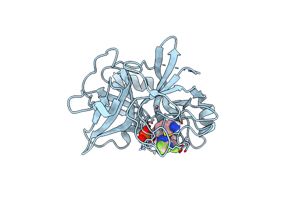
Scaffold Hopping Via Ring Opening Enables Identification Of Acyclic Compounds As New Complement Factor D Inhibitors
Organism: Homo sapiens
Method: X-RAY DIFFRACTION Resolution:2.17 Å Release Date: 2022-11-02 Classification: HYDROLASE/HYDROLASE INHIBITOR Ligands: QIE |
 |
Scaffold Hopping Via Ring Opening Enables Identification Of Acyclic Compounds As New Complement Factor D Inhibitors
Organism: Homo sapiens
Method: X-RAY DIFFRACTION Resolution:2.21 Å Release Date: 2022-11-02 Classification: HYDROLASE/HYDROLASE INHIBITOR Ligands: R7X, GOL |
 |
Scaffold Hopping Via Ring Opening Enables Identification Of Acyclic Compounds As New Complement Factor D Inhibitors
Organism: Homo sapiens
Method: X-RAY DIFFRACTION Resolution:1.99 Å Release Date: 2022-11-02 Classification: HYDROLASE/HYDROLASE INHIBITOR Ligands: S7X |
 |
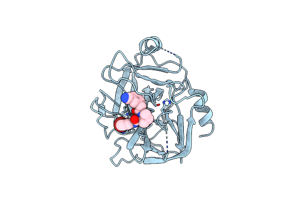
Crystal Structure Of Bcx7353(Orladeyo) In Complex With Human Plasma Kallikrein Serine Protease Domain At 2.1 Angstrom Resolution
Organism: Homo sapiens
Method: X-RAY DIFFRACTION Resolution:2.10 Å Release Date: 2021-09-15 Classification: HYDROLASE/HYDROLASE INHIBITOR Ligands: 0RI, PO4 |
 |
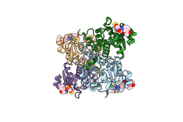
Organism: Escherichia coli o157:h7
Method: ELECTRON MICROSCOPY Release Date: 2018-12-05 Classification: RIBOSOMAL PROTEIN |
 |
Tnac-Stalled Ribosome Complex With The Titin I27 Domain Folding Close To The Ribosomal Exit Tunnel
Organism: Escherichia coli, Homo sapiens
Method: ELECTRON MICROSCOPY Release Date: 2018-12-05 Classification: RIBOSOME Ligands: MG, ZN, TRP |
 |
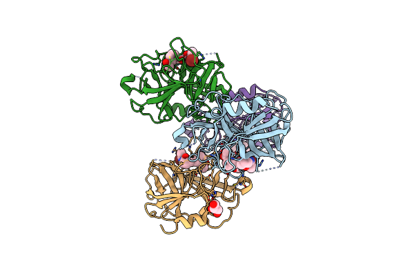
Crystal Structure Analysis Of Human Factor Viia , Souluble Tissue Factor Complexed With Bcx-3607
Organism: Homo sapiens
Method: X-RAY DIFFRACTION Resolution:2.00 Å Release Date: 2008-02-19 Classification: BLOOD CLOTTING Ligands: ASO, FUC, CA, 24X |
 |
Structure Of Ribosome Binding Domain Of The Trigger Factor On The 50S Ribosomal Subunit From D. Radiodurans
Organism: Deinococcus radiodurans
Method: X-RAY DIFFRACTION Resolution:3.35 Å Release Date: 2005-12-06 Classification: RIBOSOME |
 |
Method: X-RAY DIFFRACTION
Resolution:2.00 Å Release Date: 2001-01-10 Classification: METAL BINDING PROTEIN Ligands: CA |
 |
Method: X-RAY DIFFRACTION
Resolution:1.86 Å Release Date: 2001-01-10 Classification: METAL BINDING PROTEIN Ligands: SR |
 |
Organism: Cricket paralysis virus
Method: X-RAY DIFFRACTION Resolution:2.40 Å Release Date: 1999-08-09 Classification: VIRUS |