
Search Count: 136
 |
Organism: Methylorubrum extorquens
Method: X-RAY DIFFRACTION Release Date: 2025-10-15 Classification: TRANSPORT PROTEIN Ligands: PQQ, CL |
 |
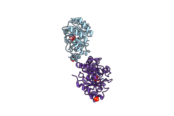
Organism: Homo sapiens, Lama glama
Method: X-RAY DIFFRACTION Resolution:2.11 Å Release Date: 2024-09-04 Classification: TRANSFERASE/INHIBITOR Ligands: EDO, XOO, SO4, ZN |
 |
Organism: Homo sapiens, Lama glama
Method: X-RAY DIFFRACTION Resolution:2.20 Å Release Date: 2024-09-04 Classification: TRANSFERASE/INHIBITOR Ligands: EDO, XOO, SO4 |
 |
Organism: Homo sapiens, Lama glama
Method: X-RAY DIFFRACTION Resolution:1.97 Å Release Date: 2024-09-04 Classification: TRANSFERASE/INHIBITOR Ligands: EDO, XOO, SO4 |
 |
Organism: Homo sapiens, Lama glama
Method: X-RAY DIFFRACTION Resolution:1.90 Å Release Date: 2024-09-04 Classification: TRANSFERASE/INHIBITOR Ligands: XQ2, EDO, SO4, ZN, CL |
 |
Organism: Homo sapiens
Method: X-RAY DIFFRACTION Resolution:2.00 Å Release Date: 2024-09-04 Classification: TRANSFERASE/INHIBITOR Ligands: XOO, EDO |
 |
Organism: Methylorubrum extorquens (strain cm4 / ncimb 13688)
Method: X-RAY DIFFRACTION Resolution:1.46 Å Release Date: 2024-08-21 Classification: TRANSPORT PROTEIN Ligands: PQQ, NA |
 |
Organism: Methylorubrum extorquens
Method: X-RAY DIFFRACTION Resolution:1.55 Å Release Date: 2024-08-21 Classification: TRANSPORT PROTEIN Ligands: EDO, PQQ, GD3 |
 |
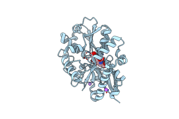
Crystal Structure Of Type I Dehydroquinase From Staphylococcus Aureus Inhibited By A Hydroxylamine Derivative
Organism: Staphylococcus aureus
Method: X-RAY DIFFRACTION Resolution:1.65 Å Release Date: 2023-02-15 Classification: LYASE Ligands: PVI, SO4, CL |
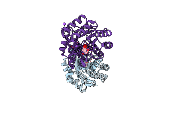 |
Crystal Structure Of Type I Dehydroquinase From Salmonella Typhi Inhibited By A Hydroxylamine Derivative
Organism: Salmonella enterica subsp. enterica serovar typhi
Method: X-RAY DIFFRACTION Resolution:1.90 Å Release Date: 2023-02-15 Classification: LYASE Ligands: PVI, NA |
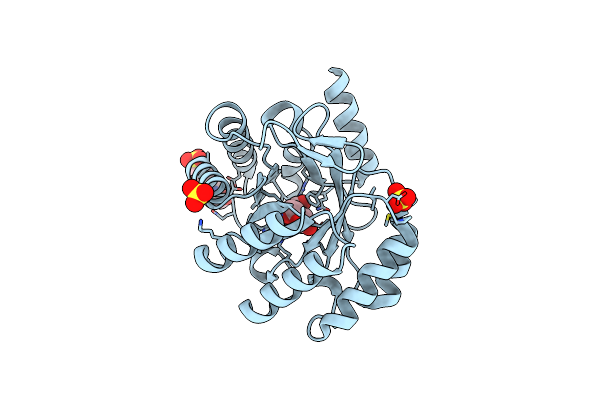 |
Crystal Structure Of Type I Dehydroquinase From Salmonella Typhi Inhibited By An Epoxide Derivative
Organism: Salmonella enterica subsp. enterica serovar typhi
Method: X-RAY DIFFRACTION Resolution:1.55 Å Release Date: 2023-02-15 Classification: LYASE Ligands: OVU, EPE, SO4 |
 |
Organism: Escherichia coli
Method: X-RAY DIFFRACTION Resolution:2.99 Å Release Date: 2022-11-23 Classification: METAL BINDING PROTEIN Ligands: X5Z, SO4, GLN, GOL |
 |
Organism: Human enterovirus d68
Method: X-RAY DIFFRACTION Resolution:1.95 Å Release Date: 2021-09-22 Classification: HYDROLASE/INHIBITOR Ligands: AG7 |
 |
Organism: Severe acute respiratory syndrome coronavirus 2
Method: X-RAY DIFFRACTION Resolution:2.10 Å Release Date: 2021-09-22 Classification: HYDROLASE/INHIBITOR Ligands: AG7 |
 |
Organism: Severe acute respiratory syndrome coronavirus 2
Method: X-RAY DIFFRACTION Resolution:2.45 Å Release Date: 2021-09-22 Classification: HYDROLASE/INHIBITOR Ligands: AG7 |
 |
Organism: Homo sapiens
Method: X-RAY DIFFRACTION Resolution:2.96 Å Release Date: 2021-06-02 Classification: HYDROLASE |
 |
Structure Of The Peptidylarginine Deiminase Type Iii (Pad3) In Complex With Cl-Amidine
Organism: Homo sapiens
Method: X-RAY DIFFRACTION Resolution:3.18 Å Release Date: 2021-06-02 Classification: HYDROLASE Ligands: BFB, CA, CL, EDO, GOL |
 |
Structure Of The Ca2+-Bound C646A Mutant Of Peptidylarginine Deiminase Type Iii (Pad3)
Organism: Homo sapiens
Method: X-RAY DIFFRACTION Resolution:3.15 Å Release Date: 2021-06-02 Classification: HYDROLASE Ligands: CA, GOL, CL |
 |
Structure Of The C646A Mutant Of Peptidylarginine Deiminase Type Iii (Pad3)
Organism: Homo sapiens
Method: X-RAY DIFFRACTION Resolution:2.10 Å Release Date: 2021-06-02 Classification: HYDROLASE Ligands: EDO, GOL |
 |
Structure Of The Inactive Form Of Wild-Type Peptidylarginine Deiminase Type Iii (Pad3) Crystallized Under The Condition With High Concentrations Of Ca2+
Organism: Homo sapiens
Method: X-RAY DIFFRACTION Resolution:2.75 Å Release Date: 2021-06-02 Classification: HYDROLASE Ligands: CA, GOL, CL |