Search Count: 13
 |
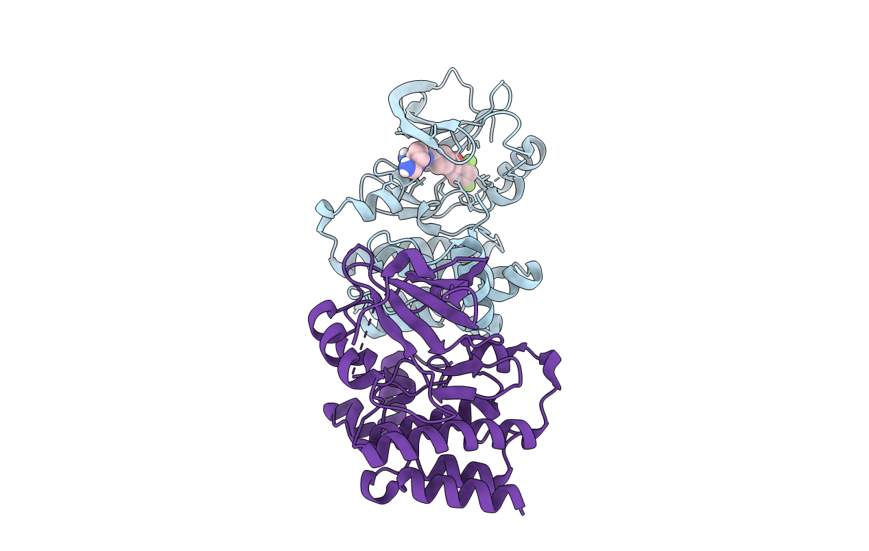
Organism: Mus musculus
Method: X-RAY DIFFRACTION Resolution:2.80 Å Release Date: 2019-01-30 Classification: TRANSFERASE Ligands: JVP |
 |
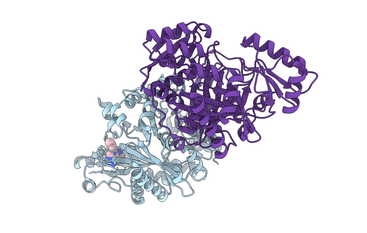
Crystal Structure Of Biotin Carboxylase From E. Coli In Complex With Amino-Oxazole Fragment Series
Organism: Escherichia coli
Method: X-RAY DIFFRACTION Resolution:2.00 Å Release Date: 2009-05-19 Classification: LIGASE Ligands: OA1, CL |
 |
Crystal Structure Of Biotin Carboxylase From E. Coli In Complex With Amino-Oxazole Fragment Series
Organism: Escherichia coli
Method: X-RAY DIFFRACTION Resolution:1.87 Å Release Date: 2009-05-19 Classification: LIGASE Ligands: OA2, CL |
 |
Crystal Structure Of Biotin Carboxylase From E. Coli In Complex With 4-Amino-7,7-Dimethyl-7,8-Dihydro-Quinazolinone Fragment
Organism: Escherichia coli
Method: X-RAY DIFFRACTION Resolution:2.50 Å Release Date: 2009-05-19 Classification: LIGASE Ligands: OA3, CL |
 |
Crystal Structure Of Biotin Carboxylase From E. Coli In Complex With 5-Methyl-6-Phenyl-Quinazoline-2,4-Diamine
Organism: Escherichia coli
Method: X-RAY DIFFRACTION Resolution:1.85 Å Release Date: 2009-05-19 Classification: LIGASE Ligands: OA4 |
 |
Crystal Structure Of Biotin Carboxylase From E. Coli In Complex With The Triazine-2,4-Diamine Fragment
Organism: Escherichia coli
Method: X-RAY DIFFRACTION Resolution:2.05 Å Release Date: 2009-05-19 Classification: LIGASE Ligands: OA5, CL |
 |
Crystal Structure Of Biotin Carboxylase From E. Coli In Complex With The 3-(3-Methyl-But-2-Enyl)-3H-Purin-6-Ylamine Fragment
Organism: Escherichia coli
Method: X-RAY DIFFRACTION Resolution:1.90 Å Release Date: 2009-05-19 Classification: LIGASE Ligands: L21, CL |
 |
Crystal Structure Of Biotin Carboxylase From E. Coli In Complex With The Amino-Thiazole-Pyrimidine Fragment
Organism: Escherichia coli
Method: X-RAY DIFFRACTION Resolution:1.77 Å Release Date: 2009-05-19 Classification: LIGASE Ligands: L22, CL |
 |
Crystal Structure Of Biotin Carboxylase From E. Coli In Complex With The Imidazole-Pyrimidine Inhibitor
Organism: Escherichia coli
Method: X-RAY DIFFRACTION Resolution:1.99 Å Release Date: 2009-05-19 Classification: LIGASE Ligands: CL, L23 |
 |
Organism: Saccharomyces cerevisiae
Method: SOLUTION NMR Release Date: 1998-04-15 Classification: TRANSCRIPTION REGULATION Ligands: CD |
 |
Nmr Solution Structure Of Growth Factor Receptor-Bound Protein 2 (Grb2) Sh2 Domain, 24 Structures
Organism: Homo sapiens
Method: SOLUTION NMR Release Date: 1997-01-27 Classification: SRC HOMOLOGY 2 DOMAIN |
 |
The Solution Structure Of Omega-Aga-Ivb, A P-Type Calcium Channel Antagonist From The Venom Of Agelenopsis Aperta
Organism: Agelenopsis aperta
Method: SOLUTION NMR Release Date: 1996-03-08 Classification: NEUROTOXIN |
 |
Solution Structure Of Fe(Ii) Cytochrome C551 From Pseudomonas Aeruginosa As Determined By Two-Dimensional 1H Nmr
Organism: Pseudomonas aeruginosa
Method: SOLUTION NMR Release Date: 1993-10-31 Classification: ELECTRON TRANSPORT Ligands: HEC |

