
Search Count: 13
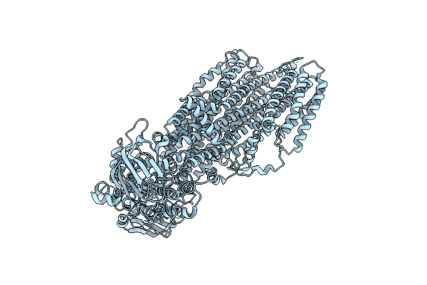 |
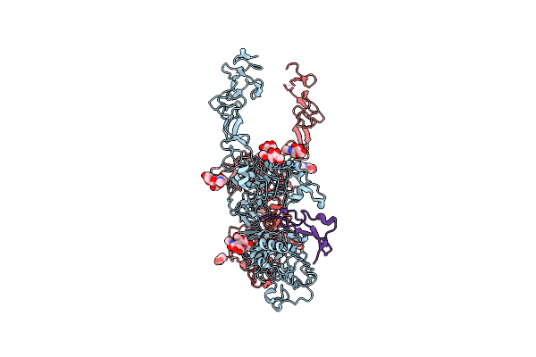
Organism: Saccharomyces cerevisiae s288c
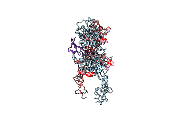
Method: ELECTRON MICROSCOPY Release Date: 2022-04-06 Classification: MEMBRANE PROTEIN |
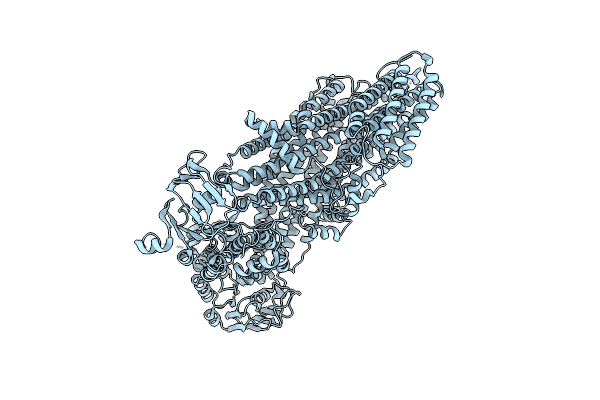 |
Organism: Saccharomyces cerevisiae s288c
Method: ELECTRON MICROSCOPY Release Date: 2022-04-06 Classification: MEMBRANE PROTEIN |
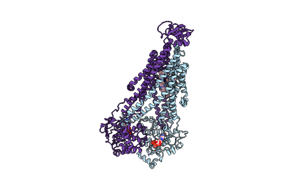 |
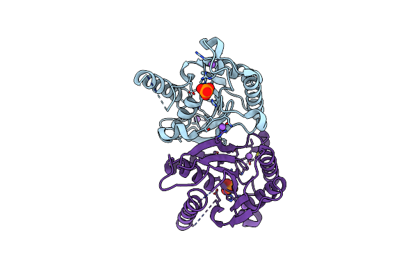
The Structure Of Bacillus Subtilis Bmrcd In The Inward-Facing Conformation Bound To Hoechst-33342 And Atp
Organism: Bacillus subtilis subsp. subtilis str. 168
Method: ELECTRON MICROSCOPY Release Date: 2022-01-05 Classification: TRANSPORT PROTEIN Ligands: ATP, HT1 |
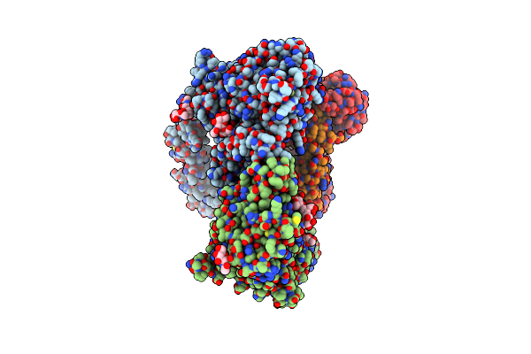 |
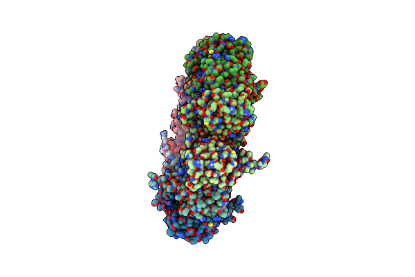
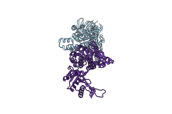
Structure Of The Her2/Her3/Nrg1B Heterodimer Extracellular Domain Bound To Trastuzumab Fab
Organism: Homo sapiens, Escherichia coli
Method: ELECTRON MICROSCOPY Release Date: 2021-11-10 Classification: SIGNALING PROTEIN/IMMUNE SYSTEM Ligands: NAG |
 |
Organism: Homo sapiens, Escherichia coli
Method: ELECTRON MICROSCOPY Release Date: 2021-10-27 Classification: SIGNALING PROTEIN Ligands: NAG |
 |
Organism: Homo sapiens, Escherichia coli
Method: ELECTRON MICROSCOPY Release Date: 2021-10-27 Classification: SIGNALING PROTEIN Ligands: NAG |
 |
Organism: Homo sapiens
Method: X-RAY DIFFRACTION Resolution:1.70 Å Release Date: 2019-02-13 Classification: SIGNALING PROTEIN |
 |
Organism: Homo sapiens
Method: X-RAY DIFFRACTION Resolution:2.60 Å Release Date: 2019-02-13 Classification: SIGNALING PROTEIN Ligands: MG |
 |
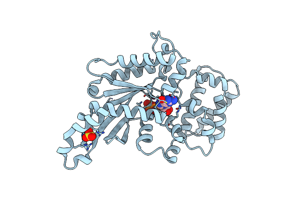
Crystal Structure Of C. Elegans Let-23 Kinase Domain Complexed With Amp-Pnp
Organism: Caenorhabditis elegans
Method: X-RAY DIFFRACTION Resolution:2.39 Å Release Date: 2018-01-31 Classification: TRANSFERASE Ligands: ANP, MG |
 |
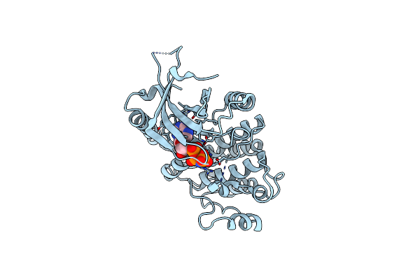
Crystal Structure Of The K345L Variant Of The Gi Alpha1 Subunit Bound To Gtpgammas
Organism: Rattus norvegicus
Method: X-RAY DIFFRACTION Resolution:1.55 Å Release Date: 2014-03-12 Classification: HYDROLASE Ligands: SO3, MG, GSP |
 |
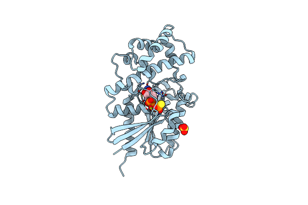
Crystal Structure Of The K345L Variant Of The Gi Alpha1 Subunit Bound To Gdp
Organism: Rattus norvegicus
Method: X-RAY DIFFRACTION Resolution:2.10 Å Release Date: 2014-03-12 Classification: HYDROLASE Ligands: GDP, SO4 |
 |
Organism: Entamoeba histolytica
Method: X-RAY DIFFRACTION Resolution:2.40 Å Release Date: 2012-12-05 Classification: TRANSFERASE |
 |
Organism: Cryptococcus neoformans
Method: X-RAY DIFFRACTION Resolution:1.89 Å Release Date: 2012-12-05 Classification: TRANSFERASE |