
Search Count: 7
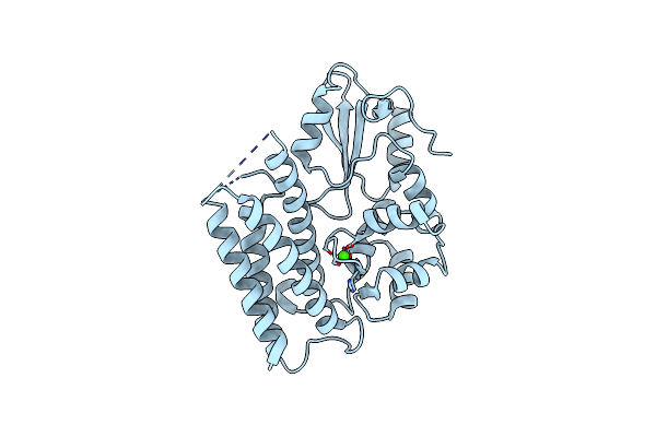 |
Organism: Homo sapiens
Method: X-RAY DIFFRACTION Resolution:1.64 Å Release Date: 2013-03-06 Classification: PROTEIN BINDING Ligands: CA |
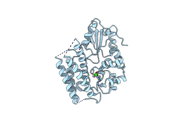 |
Crystal Structure Of The Tyrosine Kinase Binding Domain Of Cbl-C In Complex With Phospho-Src Peptide
Organism: Homo sapiens
Method: X-RAY DIFFRACTION Resolution:1.80 Å Release Date: 2013-03-06 Classification: PROTEIN BINDING/Transferase Ligands: CA |
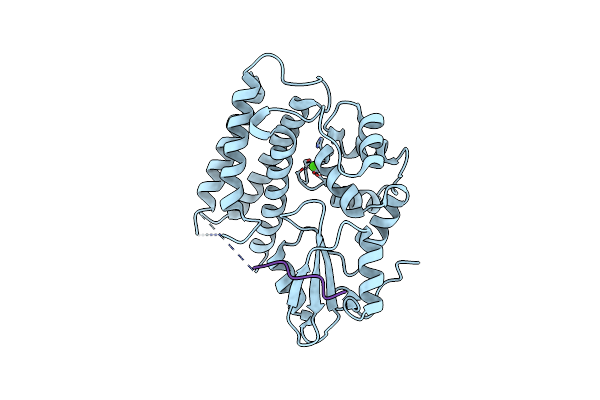 |
Crystal Structure Of The Tyrosine Kinase Binding Domain Of Cbl-C In Complex With Phospho-Egfr Peptide
Organism: Homo sapiens
Method: X-RAY DIFFRACTION Resolution:1.52 Å Release Date: 2013-03-06 Classification: PROTEIN BINDING/Transferase Ligands: CA |
 |
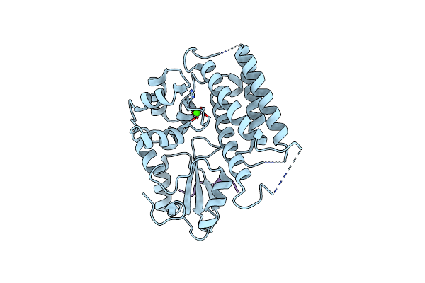
Crystal Structure Of The Tyrosine Kinase Binding Domain Of Cbl-C (Pl Mutant)
Organism: Homo sapiens
Method: X-RAY DIFFRACTION Resolution:2.39 Å Release Date: 2013-03-06 Classification: PROTEIN BINDING Ligands: CA |
 |
Crystal Structure Of The Tyrosine Kinase Binding Domain Of Cbl-C (Pl Mutant) In Complex With Phospho-Egfr Peptide
Organism: Homo sapiens
Method: X-RAY DIFFRACTION Resolution:2.00 Å Release Date: 2013-03-06 Classification: PROTEIN BINDING/TRANSFERASE Ligands: CA |
 |
 |