
Search Count: 21
 |
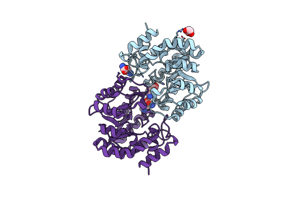
Organism: Homo sapiens
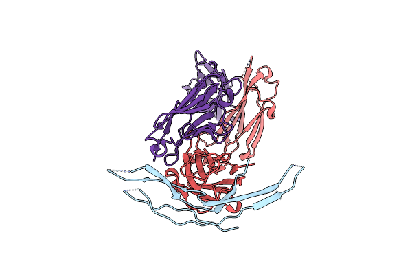
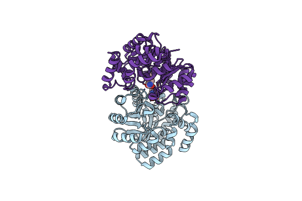
Method: X-RAY DIFFRACTION Resolution:1.95 Å Release Date: 2023-12-27 Classification: SIGNALING PROTEIN |
 |
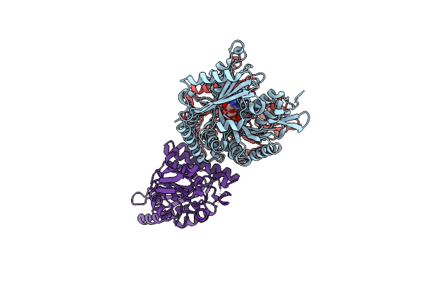
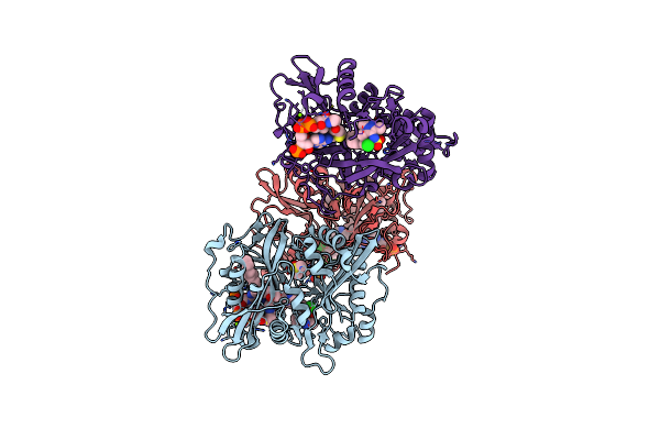
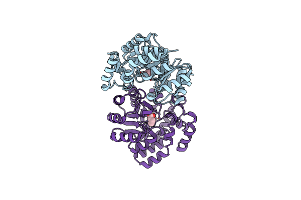
Organism: Plasmodium berghei, Sus scrofa
Method: ELECTRON MICROSCOPY Release Date: 2022-11-16 Classification: MOTOR PROTEIN Ligands: ANP, MG, GTP, G2P |
 |
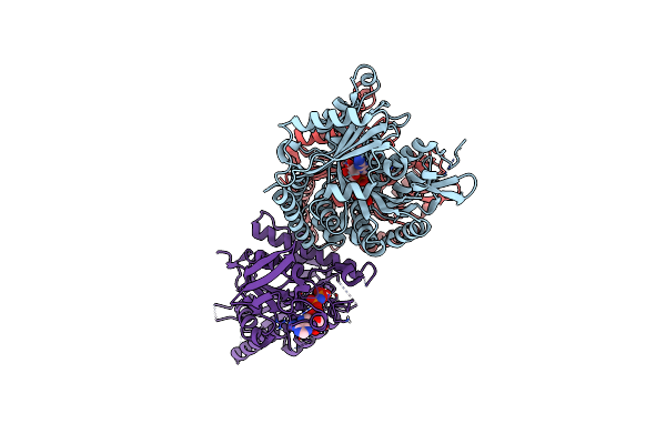
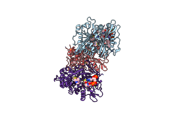
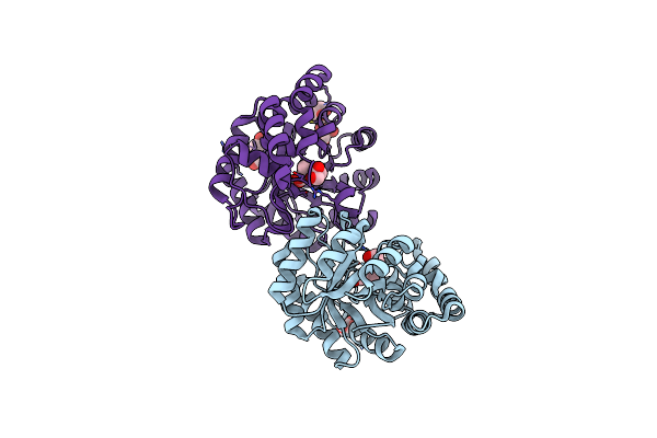
P. Berghei Kinesin-8B Motor Domain In No Nucleotide State Bound To Tubulin Dimer
Organism: Plasmodium berghei, Sus scrofa
Method: ELECTRON MICROSCOPY Release Date: 2022-10-19 Classification: MOTOR PROTEIN Ligands: GTP, MG, G2P |
 |
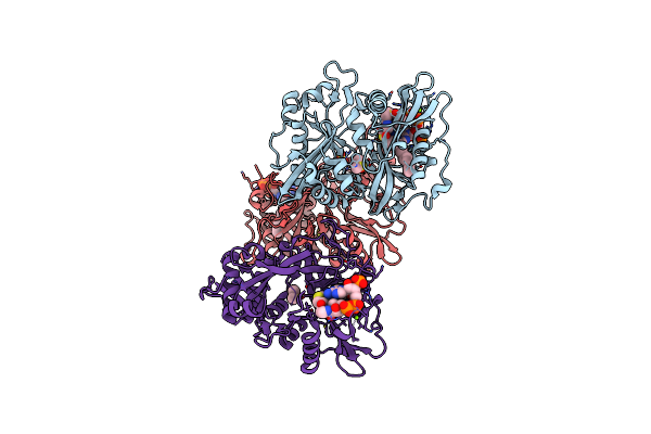
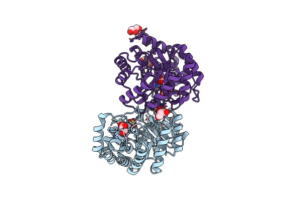
P. Falciparum Kinesin-8B Motor Domain In No Nucleotide Bound To Tubulin Dimer
Organism: Plasmodium falciparum, Sus scrofa
Method: ELECTRON MICROSCOPY Release Date: 2022-10-19 Classification: MOTOR PROTEIN Ligands: GTP, MG, G2P |
 |
Plasmodium Falciparum Kinesin-5 Motor Domain Without Nucleotide, Complexed With 14 Protofilament Microtubule.
Organism: Plasmodium falciparum (isolate 3d7), Sus scrofa
Method: ELECTRON MICROSCOPY Release Date: 2021-10-13 Classification: MOTOR PROTEIN Ligands: GTP, MG, G2P |
 |
Plasmodium Falciparum Kinesin-5 Motor Domain Bound To Amppnp, Complexed With 14 Protofilament Microtubule.
Organism: Plasmodium falciparum (isolate nf54), Sus scrofa
Method: ELECTRON MICROSCOPY Release Date: 2021-10-13 Classification: MOTOR PROTEIN Ligands: GTP, MG, G2P, ANP |
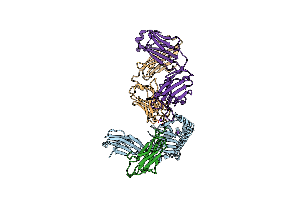 |
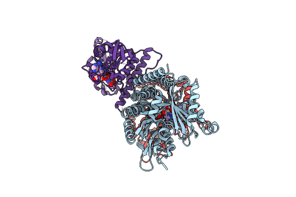
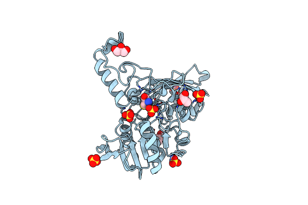
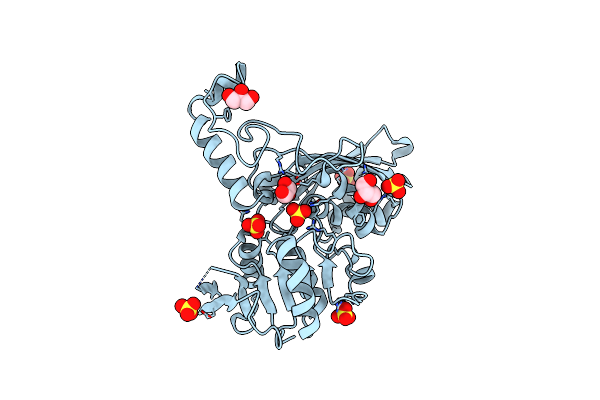
Human Fcrn Extra-Cellular Domain Complexed With Fab Fragment Of Rozanolixizumab
Organism: Homo sapiens
Method: X-RAY DIFFRACTION Resolution:2.90 Å Release Date: 2018-08-29 Classification: IMMUNE SYSTEM Ligands: CL, NA |
 |
Plasmodium Vivax N-Myristoyltransferase In Complex With Ync12-Coa Thioester.
Organism: Plasmodium vivax
Method: X-RAY DIFFRACTION Resolution:1.75 Å Release Date: 2014-01-15 Classification: TRANSFERASE Ligands: DMS, YNC, SO4, MG, CL |
 |
Plasmodium Vivax N-Myristoyltransferase In Complex With A Pyrazole Sulphonamide Inhibitor.
Organism: Plasmodium vivax
Method: X-RAY DIFFRACTION Resolution:1.89 Å Release Date: 2014-01-15 Classification: TRANSFERASE Ligands: DMS, NHW, 646, SO4, MG, CL |
 |
Plasmodium Vivax N-Myristoyltransferase In Complex With A Benzothiophene Inhibitor
Organism: Plasmodium vivax
Method: X-RAY DIFFRACTION Resolution:1.72 Å Release Date: 2014-01-15 Classification: TRANSFERASE Ligands: DMS, NHW, YNE, SO4, MG, CL |
 |
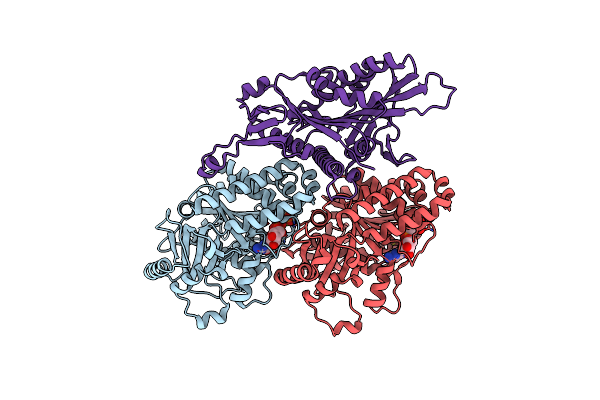
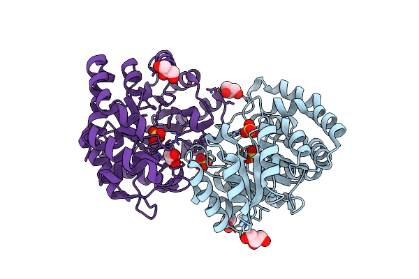
Crystal Structure Of Aspartate Semialdehyde Dehydrogenase Complexed With Inhibitor Smcs (Cys) And Phosphate From Mycobacterium Tuberculosis H37Rv
Organism: Mycobacterium tuberculosis
Method: X-RAY DIFFRACTION Resolution:1.95 Å Release Date: 2012-05-30 Classification: OXIDOREDUCTASE Ligands: CYS, GOL, SO4 |
 |
Crystal Structure Of Aspartate Semialdehyde Dehydrogenase Complexed With Glycerol And Sulfate From Mycobacterium Tuberculosis H37Rv
Organism: Mycobacterium tuberculosis
Method: X-RAY DIFFRACTION Resolution:2.18 Å Release Date: 2012-05-30 Classification: OXIDOREDUCTASE Ligands: GOL, SO4 |
 |
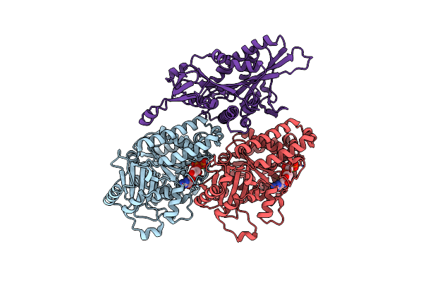
Crystal Structure Of The Complex Of Dihydrodipicolinate Synthase From Acinetobacter Baumannii With 5-Hydroxylysine At 2.6 A Resolution
Organism: Acinetobacter baumannii
Method: X-RAY DIFFRACTION Resolution:2.60 Å Release Date: 2011-08-31 Classification: LYASE Ligands: LYZ |
 |
Crystal Structure Of The Complex Of Dihydrodipicolinate Synthase From Acinetobacter Baumannii With 2-Ketobutanoic Acid At 1.99 A Resolution
Organism: Acinetobacter baumannii
Method: X-RAY DIFFRACTION Resolution:1.99 Å Release Date: 2011-08-31 Classification: LYASE Ligands: 2KT |
 |
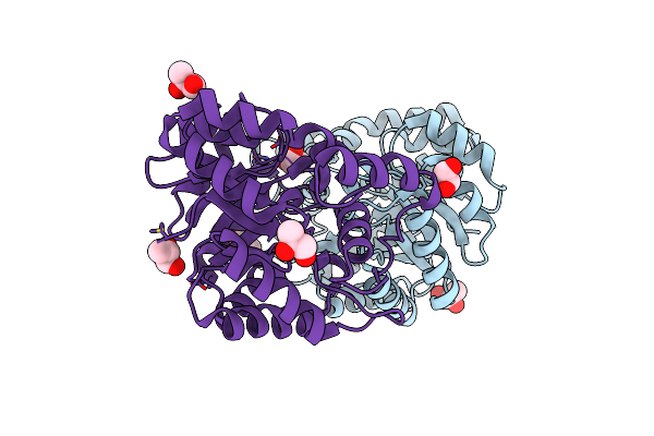
Crystal Structure Of The Complex Of Dhdps From Acinetobacter Baumannii With Pyruvate At 1.4 A Resolution
Organism: Acinetobacter baumannii
Method: X-RAY DIFFRACTION Resolution:1.42 Å Release Date: 2011-08-24 Classification: LYASE Ligands: GOL, PYR, PEG |
 |
Crystal Structure Of The Complex Of Dhydrodipicolinate Synthase From Acinetobacter Baumannii With Lysine At 2.3A Resolution
Organism: Acinetobacter baumannii
Method: X-RAY DIFFRACTION Resolution:2.30 Å Release Date: 2010-12-29 Classification: LYASE Ligands: GOL, SO4, LYS, ACT |
 |
Crystal Structure Of Dihydrodipicolinate Synthase From Pseudomonas Aeruginosa(Psdhdps)Complexed With L-Lysine At 2.65A Resolution
Organism: Pseudomonas aeruginosa
Method: X-RAY DIFFRACTION Resolution:2.65 Å Release Date: 2010-12-29 Classification: LYASE Ligands: LYS, GOL |
 |
Crystal Structure Of Dhydrodipicolinate Synthase From Acinetobacter Baumannii At 2.8A Resolution
Organism: Acinetobacter baumannii
Method: X-RAY DIFFRACTION Resolution:2.80 Å Release Date: 2010-12-22 Classification: LYASE Ligands: SO4, GOL |
 |
Crystal Structure Of The Complex Of Dhydrodipicolinate Synthase From Acinetobacter Baumannii With Lysine At 2.6A Resolution
Organism: Acinetobacter baumannii
Method: X-RAY DIFFRACTION Resolution:2.60 Å Release Date: 2010-12-22 Classification: LYASE Ligands: SO4, LYS, GOL |
 |
Biochemical Studies And Crystal Structure Determination Of Dihydrodipicolinate Synthase From Pseudomonas Aeruginosa
Organism: Pseudomonas aeruginosa
Method: X-RAY DIFFRACTION Resolution:2.85 Å Release Date: 2010-12-15 Classification: LYASE Ligands: PGO |