
Search Count: 19
 |
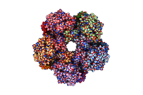
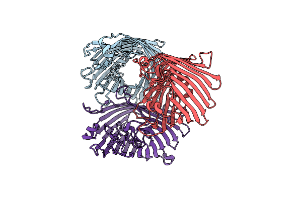
Mosquitocidal Cry11Aa Determined At Ph 7 From Naturally-Occurring Nanocrystals By Serial Femtosecond Crystallography
Organism: Bacillus thuringiensis serovar israelensis
Method: X-RAY DIFFRACTION Resolution:2.60 Å Release Date: 2022-07-27 Classification: TOXIN |
 |
Mosquitocidal Cry11Aa-Y449F Determined At Ph 7 From Naturally-Occurring Nanocrystals By Serial Femtosecond Crystallography
Organism: Bacillus thuringiensis serovar israelensis
Method: X-RAY DIFFRACTION Resolution:3.10 Å Release Date: 2022-07-27 Classification: TOXIN |
 |
Mosquitocidal Cry11Aa-E583Q Determined At Ph 7 From Naturally-Occurring Nanocrystals By Serial Femtosecond Crystallography
Organism: Bacillus thuringiensis serovar israelensis
Method: X-RAY DIFFRACTION Resolution:3.30 Å Release Date: 2022-07-27 Classification: TOXIN |
 |
Mosquitocidal Cry11Aa-F17Y Determined At Ph 7 From Naturally-Occurring Nanocrystals By Serial Femtosecond Crystallography
Organism: Bacillus thuringiensis serovar israelensis
Method: X-RAY DIFFRACTION Resolution:3.40 Å Release Date: 2022-07-27 Classification: TOXIN |
 |
Mosquitocidal Cry11Ba Determined At Ph 6.5 From Naturally-Occurring Nanocrystals By Serial Femtosecond Crystallography
Organism: Bacillus thuringiensis serovar jegathesan
Method: X-RAY DIFFRACTION Resolution:2.40 Å Release Date: 2022-07-27 Classification: TOXIN |
 |
Mosquitocidal Cry11Ba Determined At Ph 10.4 From Naturally-Occurring Nanocrystals By Serial Femtosecond Crystallography
Organism: Bacillus thuringiensis serovar jegathesan
Method: X-RAY DIFFRACTION Resolution:2.65 Å Release Date: 2022-07-27 Classification: TOXIN Ligands: GOL |
 |
Organism: Providencia stuartii
Method: ELECTRON MICROSCOPY Release Date: 2022-04-20 Classification: LYASE |
 |
Organism: Providencia stuartii
Method: ELECTRON MICROSCOPY Release Date: 2022-04-20 Classification: LYASE |
 |
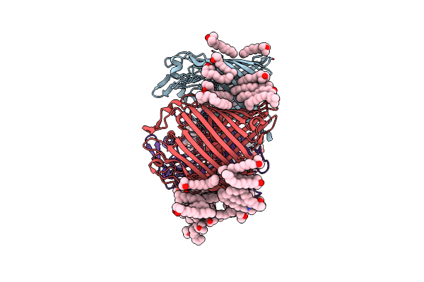
Native Structure Of Mosquitocidal Cyt1A Protoxin Obtained By Serial Femtosecond Crystallography On In Vivo Grown Crystals At Ph 7
Organism: Bacillus thuringiensis subsp. israelensis
Method: X-RAY DIFFRACTION Resolution:1.86 Å Release Date: 2020-10-14 Classification: TOXIN |
 |
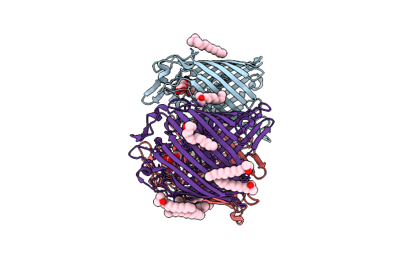
Structure Of Mosquitocidal Cyt1A Protoxin Obtained By Serial Femtosecond Crystallography On In Vivo Grown Crystals Soaked With Dtt At Ph 7
Organism: Bacillus thuringiensis subsp. israelensis
Method: X-RAY DIFFRACTION Resolution:1.85 Å Release Date: 2020-10-14 Classification: TOXIN |
 |
Structure Of Mosquitocidal Cyt1Aa Protoxin Obtained By Serial Femtosecond Crystallography On In Vivo Grown Crystals At Ph 10
Organism: Bacillus thuringiensis subsp. israelensis
Method: X-RAY DIFFRACTION Resolution:1.85 Å Release Date: 2020-10-14 Classification: TOXIN Ligands: CA |
 |
Structure Of The C7S Mutant Of Mosquitocidal Cyt1A Protoxin Obtained By Serial Femtosecond Crystallography On In Vivo Grown Crystals At Ph 7
Organism: Bacillus thuringiensis subsp. israelensis
Method: X-RAY DIFFRACTION Resolution:2.00 Å Release Date: 2020-10-14 Classification: TOXIN |
 |
Structure Of 283-Lgny-286, The Steric Zipper That Supports The Self-Association Of P. Stuartii Omp-Pst2 Into Dimers Of Trimers
Organism: Providencia stuartii
Method: X-RAY DIFFRACTION Resolution:1.00 Å Release Date: 2018-02-21 Classification: CELL ADHESION Ligands: SO4 |
 |
Structure Of 206-Gvvtse-211, The Steric Zipper That Supports The Self-Association Of P. Stuartii Omp-Pst1 Into Dimers Of Trimers
Organism: Providencia stuartii
Method: X-RAY DIFFRACTION Resolution:1.91 Å Release Date: 2018-02-21 Classification: CELL ADHESION |
 |
Organism: Providencia stuartii
Method: X-RAY DIFFRACTION Resolution:3.12 Å Release Date: 2018-02-21 Classification: TRANSPORT PROTEIN Ligands: CA |
 |
Organism: Providencia stuartii
Method: X-RAY DIFFRACTION Resolution:2.70 Å Release Date: 2018-02-21 Classification: CELL ADHESION Ligands: LDA, CA, CL |
 |
Organism: Providencia stuartii
Method: X-RAY DIFFRACTION Resolution:3.00 Å Release Date: 2018-02-21 Classification: CELL ADHESION Ligands: LDA, CA |
 |
Structure Of Porin Omp-Pst1 From P. Stuartii; The Crystallographic Symmetry Generates A Dimer Of Trimers.
Organism: Providencia stuartii
Method: X-RAY DIFFRACTION Resolution:3.20 Å Release Date: 2016-03-09 Classification: TRANSPORT PROTEIN Ligands: CA |
 |
Structure Of Porin Omp-Pst2 From P. Stuartii; The Asymmetric Unit Contains A Dimer Of Trimers.
Organism: Providencia stuartii
Method: X-RAY DIFFRACTION Resolution:2.20 Å Release Date: 2016-03-09 Classification: TRANSPORT PROTEIN Ligands: LDA, FTT, MYR, SO4 |