
Search Count: 16
 |
Crystal Structure Of Acinetobacter Sp. Dl28 L-Ribose Isomerase In Complex With L-Ribose
Organism: Acinetobacter
Method: X-RAY DIFFRACTION Resolution:1.93 Å Release Date: 2014-05-28 Classification: ISOMERASE Ligands: CO, 0MK, Z6J, NCO |
 |
Crystal Structure Of Acinetobacter Sp. Dl28 L-Ribose Isomerase In Complex With L-Ribulose
Organism: Acinetobacter
Method: X-RAY DIFFRACTION Resolution:1.93 Å Release Date: 2014-05-28 Classification: ISOMERASE Ligands: CO, RUU, QDK, NCO |
 |
Crystal Structure Of Acinetobacter Sp. Dl28 L-Ribose Isomerase In Complex With Ribitol
Organism: Acinetobacter
Method: X-RAY DIFFRACTION Resolution:1.93 Å Release Date: 2014-05-28 Classification: ISOMERASE Ligands: CO, RB0, NCO |
 |
Crystal Structure Of Acinetobacter Sp. Dl28 L-Ribose Isomerase Mutant E204Q In Complex With L-Ribose
Organism: Acinetobacter
Method: X-RAY DIFFRACTION Resolution:1.98 Å Release Date: 2014-05-28 Classification: ISOMERASE Ligands: CO, ROR, Z6J, 0MK, NCO |
 |
Crystal Structure Of Acinetobacter Sp. Dl28 L-Ribose Isomerase Mutant E204Q In Complex With L-Ribulose
Organism: Acinetobacter
Method: X-RAY DIFFRACTION Resolution:1.98 Å Release Date: 2014-05-28 Classification: ISOMERASE Ligands: CO, QDK, RUU, NCO |
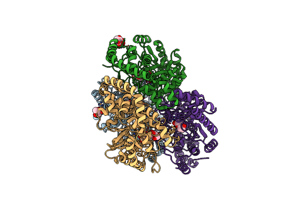 |
Crystal Structure Of Pseudomonas Stutzeri L-Rhamnose Isomerase Mutant H101N In Complex With L-Rhamnopyranose
Organism: Pseudomonas stutzeri
Method: X-RAY DIFFRACTION Resolution:1.70 Å Release Date: 2012-12-12 Classification: ISOMERASE Ligands: RNS, RAM, MN, RM4 |
 |
Crystal Structure Of Pseudomonas Stutzeri L-Rhamnose Isomerase Mutant H101N In Complex With D-Allopyranose
Organism: Pseudomonas stutzeri
Method: X-RAY DIFFRACTION Resolution:2.38 Å Release Date: 2012-12-12 Classification: ISOMERASE Ligands: MN, AOS, AFD |
 |
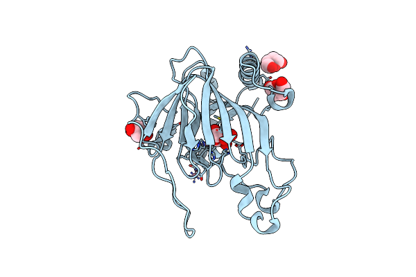
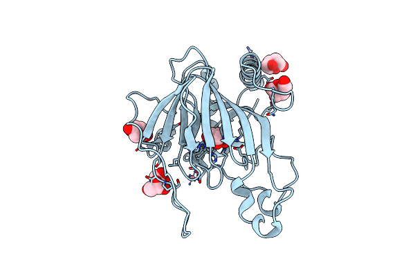
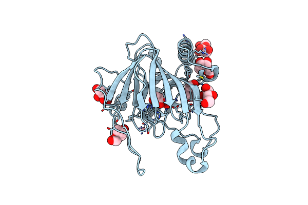
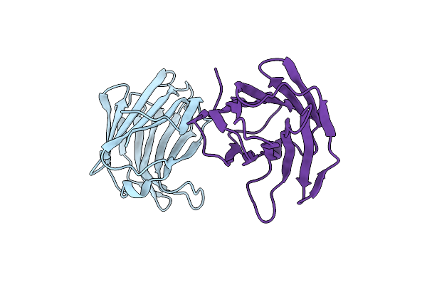
Protease-Resistant Mutant Form Of Human Galectin-8 In Complex With Two Lactose Molecules
Organism: Homo sapiens
Method: X-RAY DIFFRACTION Resolution:2.55 Å Release Date: 2012-09-12 Classification: SUGAR BINDING PROTEIN Ligands: EDO |
 |
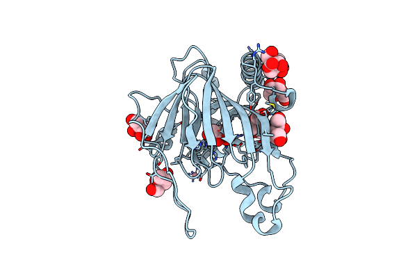
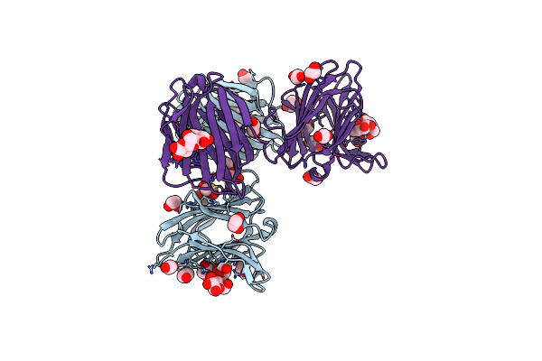
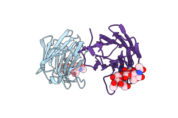
Protease-Resistant Mutant Form Of Human Galectin-8 In Complex With Sialyllactose And Lactose
Organism: Homo sapiens
Method: X-RAY DIFFRACTION Resolution:2.98 Å Release Date: 2012-09-12 Classification: SUGAR BINDING PROTEIN Ligands: EDO |
 |
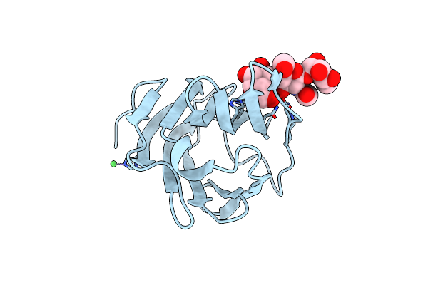
Organism: Homo sapiens
Method: X-RAY DIFFRACTION Resolution:1.98 Å Release Date: 2012-09-12 Classification: SUGAR BINDING PROTEIN Ligands: CL |
 |
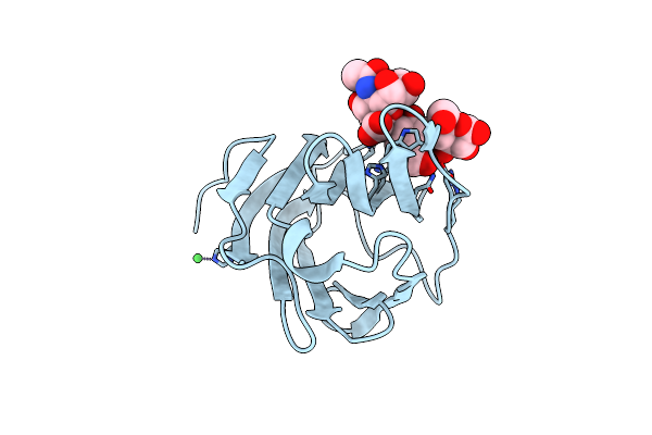
Organism: Homo sapiens
Method: X-RAY DIFFRACTION Resolution:2.08 Å Release Date: 2012-09-12 Classification: SUGAR BINDING PROTEIN Ligands: CL |
 |
Organism: Homo sapiens
Method: X-RAY DIFFRACTION Resolution:2.80 Å Release Date: 2012-07-11 Classification: SUGAR BINDING PROTEIN Ligands: EDO |
 |
Organism: Homo sapiens
Method: X-RAY DIFFRACTION Resolution:1.50 Å Release Date: 2010-09-22 Classification: SUGAR BINDING PROTEIN Ligands: NI |
 |
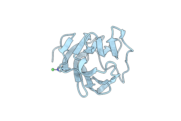
Crystal Structure Of Human Galectin-9 C-Terminal Crd In Complex With N-Acetyllactosamine
Organism: Homo sapiens
Method: X-RAY DIFFRACTION Resolution:2.34 Å Release Date: 2010-09-22 Classification: SUGAR BINDING PROTEIN Ligands: NI |
 |
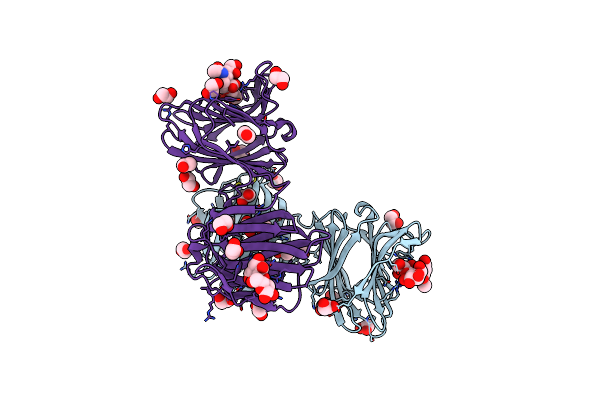
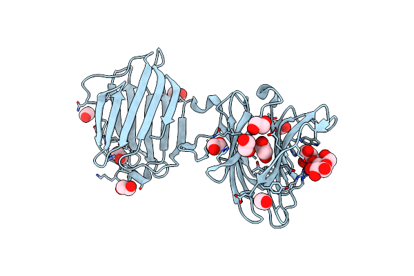
Crystal Structure Of Human Galectin-9 C-Terminal Crd In Complex With Biantennary Oligosaccharide
Organism: Homo sapiens
Method: X-RAY DIFFRACTION Resolution:1.57 Å Release Date: 2010-09-22 Classification: SUGAR BINDING PROTEIN Ligands: NI |
 |
Crystal Structure Of Human Galectin-9 C-Terminal Crd In Complex With Sialyllactose
Organism: Homo sapiens
Method: X-RAY DIFFRACTION Resolution:1.99 Å Release Date: 2010-09-22 Classification: SUGAR BINDING PROTEIN Ligands: NI |