
Search Count: 40
 |
Organism: Severe acute respiratory syndrome coronavirus 2, Tequatrovirus t4, Homo sapiens
Method: ELECTRON MICROSCOPY Release Date: 2025-07-23 Classification: PROTEIN BINDING Ligands: NAG |
 |
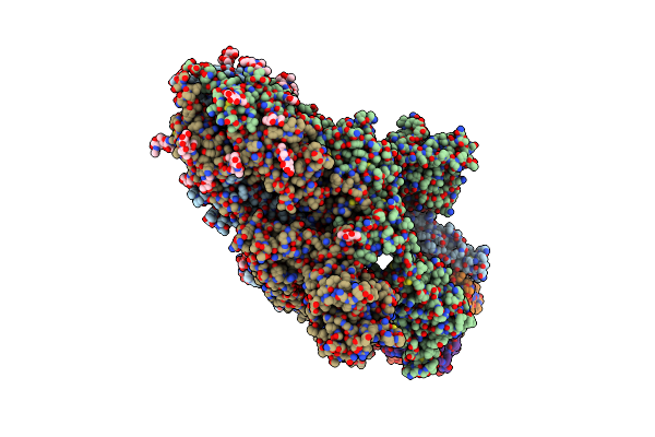
Cryo-Em Structure Of Hku25-Batcov S-Trimer Stabilized With 2P And X1 Disulfide Bond
Organism: Hypsugo bat coronavirus hku25, Tequatrovirus t4
Method: ELECTRON MICROSCOPY Release Date: 2025-06-18 Classification: VIRAL PROTEIN Ligands: EIC, NAG |
 |
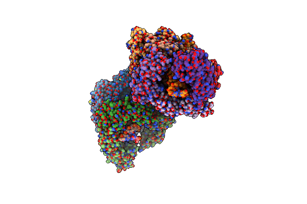
Cryo-Em Structure Of Cn-Hedgehogcov (Hku31/Erinaceus Amurensis/China/2014) S-Trimer In A Locked-1 Conformation
Organism: Erinaceus hedgehog coronavirus hku31, Tequatrovirus t4
Method: ELECTRON MICROSCOPY Release Date: 2025-06-18 Classification: VIRAL PROTEIN Ligands: NAG, FOL, EIC |
 |
Organism: Bat coronavirus, Tequatrovirus t4
Method: ELECTRON MICROSCOPY Release Date: 2025-06-18 Classification: VIRAL PROTEIN Ligands: NAG, EIC |
 |
Organism: Middle east respiratory syndrome-related coronavirus, Tequatrovirus t4
Method: ELECTRON MICROSCOPY Release Date: 2025-06-18 Classification: VIRAL PROTEIN Ligands: NAG, EIC |
 |
Organism: Tequatrovirus t4
Method: X-RAY DIFFRACTION Release Date: 2025-05-14 Classification: VIRAL PROTEIN |
 |
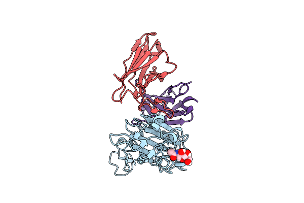
Sars-Cov-2 Xbb.1.16 Spike In Complex With Cyfn1006-1(S-Cyfn1006-1 Dimer Trimer).
Organism: Severe acute respiratory syndrome coronavirus 2, Tequatrovirus t4, Synthetic construct, Homo sapiens
Method: ELECTRON MICROSCOPY Release Date: 2025-02-12 Classification: VIRAL PROTEIN/IMMUNE SYSTEM |
 |
Structure Of Xbb.1.16 S Trimer With 2 Down-Rbds Complex With Antibody Cyfn1006-1.
Organism: Homo sapiens, Severe acute respiratory syndrome coronavirus 2, Tequatrovirus t4, Synthetic construct
Method: ELECTRON MICROSCOPY Release Date: 2025-02-12 Classification: VIRAL PROTEIN/IMMUNE SYSTEM |
 |
Sars-Cov-2 Eg.5.1 Spike In Complex With Cyfn1006-2(S-Cyfn1006-2 Dimer Trimer).
Organism: Severe acute respiratory syndrome coronavirus 2, Tequatrovirus t4, Synthetic construct, Homo sapiens
Method: ELECTRON MICROSCOPY Release Date: 2025-02-12 Classification: VIRAL PROTEIN/IMMUNE SYSTEM |
 |
Structure Of Eg.5.1 S Trimer With 3 Down-Rbds Complex With Antibody Cyfn1006-2.
Organism: Severe acute respiratory syndrome coronavirus 2, Tequatrovirus t4, Synthetic construct, Homo sapiens
Method: ELECTRON MICROSCOPY Release Date: 2025-02-12 Classification: VIRAL PROTEIN/IMMUNE SYSTEM |
 |
Structure Of Xbb.1.16 S Trimer With 3 Down-Rbds Complex With Antibody Cyfn1006-1.
Organism: Severe acute respiratory syndrome coronavirus 2, Tequatrovirus t4, Synthetic construct, Homo sapiens
Method: ELECTRON MICROSCOPY Release Date: 2025-01-29 Classification: VIRAL PROTEIN/IMMUNE SYSTEM |
 |
Organism: Severe acute respiratory syndrome coronavirus 2, Tequatrovirus t4, Synthetic construct, Homo sapiens
Method: ELECTRON MICROSCOPY Release Date: 2025-01-29 Classification: VIRAL PROTEIN/IMMUNE SYSTEM |
 |
Organism: Severe acute respiratory syndrome coronavirus 2, Tequatrovirus t4, Synthetic construct, Homo sapiens
Method: ELECTRON MICROSCOPY Release Date: 2025-01-29 Classification: VIRAL PROTEIN/IMMUNE SYSTEM |
 |
Organism: Severe acute respiratory syndrome coronavirus 2, Tequatrovirus t4, Synthetic construct, Homo sapiens
Method: ELECTRON MICROSCOPY Release Date: 2025-01-22 Classification: VIRAL PROTEIN |
 |
Computational Design Of Highly Signaling Active Membrane Receptors Through De Novo Solvent-Mediated Allosteric Networks
Organism: Tequatrovirus t4, Homo sapiens
Method: X-RAY DIFFRACTION Resolution:3.90 Å Release Date: 2024-12-18 Classification: MEMBRANE PROTEIN Ligands: NGI |
 |
Structure Of A Sars-Cov-2 Spike S2 Subunit In A Pre-Fusion, Open Conformation
Organism: Severe acute respiratory syndrome coronavirus 2, Tequatrovirus t4, Homo sapiens
Method: ELECTRON MICROSCOPY Release Date: 2024-12-18 Classification: VIRAL PROTEIN Ligands: NAG |
 |
Stabilised Ba.1 Sars-Cov-2 Spike With H6 Nanobodies In '3 Up' Rbd Conformation
Organism: Severe acute respiratory syndrome coronavirus 2, Tequatrovirus t4, Lama glama
Method: ELECTRON MICROSCOPY Release Date: 2024-07-03 Classification: VIRAL PROTEIN Ligands: NAG |
 |
Organism: Severe acute respiratory syndrome coronavirus 2, Tequatrovirus t4, Homo sapiens
Method: ELECTRON MICROSCOPY Release Date: 2024-05-08 Classification: VIRAL PROTEIN |
 |
Organism: Severe acute respiratory syndrome coronavirus 2, Tequatrovirus t4, Homo sapiens
Method: ELECTRON MICROSCOPY Release Date: 2024-01-31 Classification: VIRAL PROTEIN |
 |
Structure Of T4 Bacteriophage Clamp Loader Mutant D110C Bound To The T4 Clamp, Primer-Template Dna, And Atp Analog
Organism: Tequatrovirus t4
Method: X-RAY DIFFRACTION Resolution:3.10 Å Release Date: 2023-12-13 Classification: DNA BINDING PROTEIN/DNA Ligands: AF3, ADP, MG |