
Search Count: 10
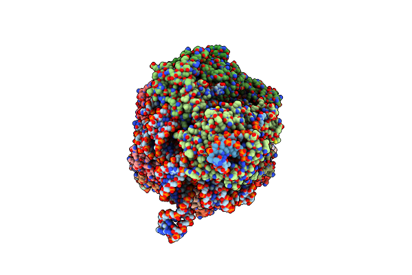 |
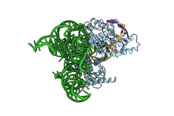
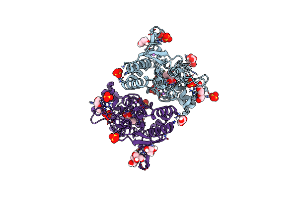
Conformational Landscape Of The Type V-K Crispr-Associated Transposonintegration Assembly Cast V-K Composite Map
Organism: Scytonema hofmannii
Method: ELECTRON MICROSCOPY Release Date: 2024-06-19 Classification: DNA BINDING PROTEIN Ligands: MG, ZN, ATP |
 |
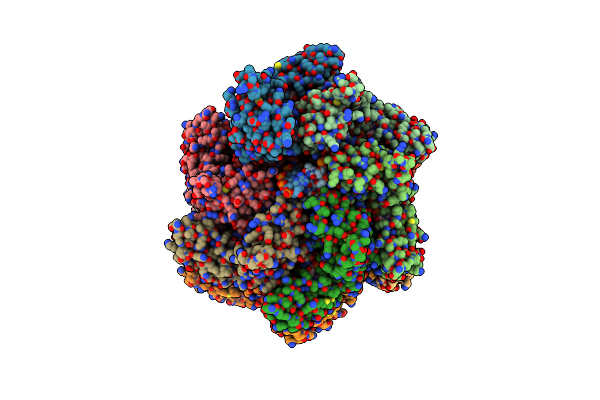
Conformational Landscape Of The Type V-K Crispr-Associated Transposonintegration Assembly Cast V-K Cas12K Domain Local-Refinement Map
Organism: Scytonema hofmannii
Method: ELECTRON MICROSCOPY Release Date: 2024-06-19 Classification: DNA BINDING PROTEIN Ligands: MG, ZN |
 |
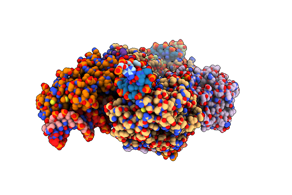
Conformational Landscape Of The Type V-K Crispr-Associated Transposonintegration Assembly Cast V-K Tnsc Domain Local-Refinement Map
Organism: Scytonema hofmannii
Method: ELECTRON MICROSCOPY Release Date: 2024-06-19 Classification: DNA BINDING PROTEIN Ligands: MG, ATP |
 |
Conformational Landscape Of The Type V-K Crispr-Associated Transposonintegration Assembly Cast V-K Tnsb Domain Local-Refinement Map
Organism: Scytonema hofmannii
Method: ELECTRON MICROSCOPY Release Date: 2024-06-19 Classification: DNA BINDING PROTEIN Ligands: MG |
 |
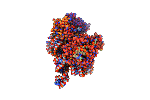
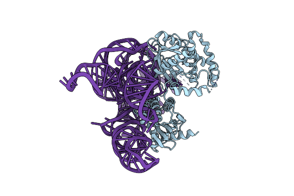
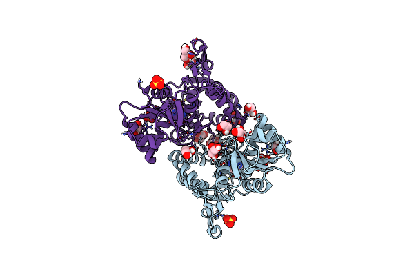
Cryo-Em Structure Of Shcas12K-Sgrna-Dsdna Ternary Complex (Type V-K Crispr-Associated Transposon)
Organism: Scytonema hofmannii
Method: ELECTRON MICROSCOPY Release Date: 2024-04-10 Classification: DNA BINDING PROTEIN |
 |
Cryo-Em Structure Of Cas12K-Sgrna Binary Complex (Type V-K Crispr-Associated Transposon)
Organism: Scytonema hofmannii
Method: ELECTRON MICROSCOPY Release Date: 2024-04-10 Classification: DNA BINDING PROTEIN |
 |
Organism: Phage #d, Synthetic construct
Method: ELECTRON MICROSCOPY Release Date: 2021-07-21 Classification: RNA BINDING PROTEIN Ligands: NI, ZN |
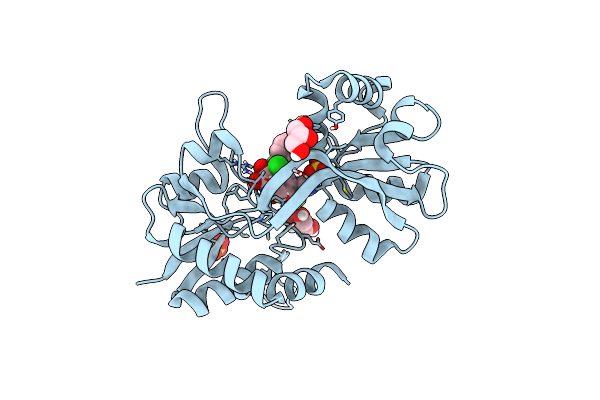 |
Structure Of Gluk1 Ligand-Binding Domain (S1S2) In Complex With N-(7-(1H-Imidazol-1-Yl)-2,3-Dioxo-6-(Trifluoromethyl)-3,4-Dihydroquinoxalin-1(2H)-Yl Benzamide At 2.3 A Resolution
Organism: Rattus norvegicus
Method: X-RAY DIFFRACTION Resolution:2.30 Å Release Date: 2019-10-30 Classification: MEMBRANE PROTEIN Ligands: SO4, L5H, CL, GOL |
 |
Structure Of Glua2 Ligand-Binding Domain (S1S2J) In Complex With The Agonist (S)-2-Amino-3-(1-Ethyl-4-Hydroxy-1H-1,2,3-Triazol-5-Yl)Propanoic Acid At 1.4 A Resolution
Organism: Rattus norvegicus
Method: X-RAY DIFFRACTION Resolution:1.40 Å Release Date: 2019-04-17 Classification: MEMBRANE PROTEIN Ligands: SO4, HJ8, GOL, PGE, CL, CIT, LI, PEG |
 |
Structure Of Glua2 Ligand-Binding Domain (S1S2J) In Complex With The Agonist (S)-2-Amino-3-(2-Methyl-5-Hydroxy-2H-1,2,3-Triazol-4-Yl)Propanoic Acid At 1.55 A Resolution
Organism: Rattus norvegicus
Method: X-RAY DIFFRACTION Resolution:1.55 Å Release Date: 2019-04-17 Classification: MEMBRANE PROTEIN Ligands: HJH, SO4, GOL, CL, LI |