
Search Count: 47
 |
Organism: Homo sapiens, Synthetic construct
Method: ELECTRON MICROSCOPY Release Date: 2025-10-22 Classification: MEMBRANE PROTEIN |
 |
Organism: Trypanosoma brucei brucei
Method: ELECTRON MICROSCOPY Release Date: 2024-10-02 Classification: MEMBRANE PROTEIN Ligands: VO6 |
 |
Organism: Trypanosoma brucei brucei
Method: ELECTRON MICROSCOPY Release Date: 2024-10-02 Classification: MEMBRANE PROTEIN Ligands: PNT |
 |
Organism: Trypanosoma brucei brucei
Method: ELECTRON MICROSCOPY Release Date: 2024-10-02 Classification: MEMBRANE PROTEIN Ligands: GOL |
 |
Structure Of The Murine Trace Amine-Associated Receptor Taar7F Bound To N,N-Dimethylcyclohexylamine (Dmch) In Complex With Mini-Gs Trimeric G Protein
Organism: Homo sapiens, Lama glama, Mus musculus
Method: ELECTRON MICROSCOPY Release Date: 2023-08-09 Classification: MEMBRANE PROTEIN Ligands: Y01, 8IA |
 |
Organism: Saccharomyces cerevisiae
Method: ELECTRON MICROSCOPY Release Date: 2022-03-16 Classification: MEMBRANE PROTEIN Ligands: NAG, Y01 |
 |
Organism: Saccharomyces cerevisiae
Method: ELECTRON MICROSCOPY Release Date: 2022-03-16 Classification: MEMBRANE PROTEIN Ligands: NAG, Y01 |
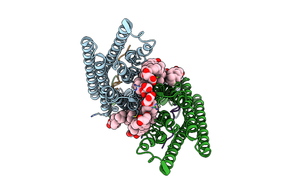 |
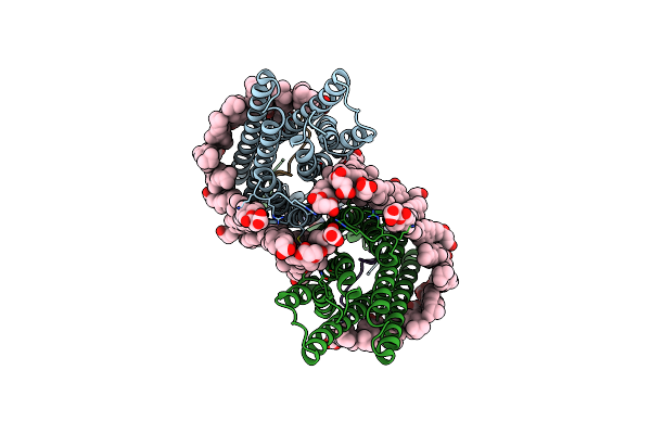
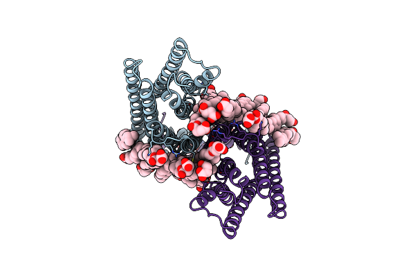
Structure Of The Gpcr Dimer Ste2 In The Inactive-Like State Bound To Agonist
Organism: Saccharomyces cerevisiae
Method: ELECTRON MICROSCOPY Release Date: 2022-03-16 Classification: MEMBRANE PROTEIN Ligands: NAG, Y01 |
 |
Organism: Saccharomyces cerevisiae
Method: ELECTRON MICROSCOPY Release Date: 2022-03-16 Classification: MEMBRANE PROTEIN Ligands: NAG, Y01 |
 |
Organism: Saccharomyces cerevisiae, Saccharomyces cerevisiae (strain atcc 204508 / s288c)
Method: ELECTRON MICROSCOPY Release Date: 2020-12-09 Classification: MEMBRANE PROTEIN Ligands: NAG, Y01 |
 |
Phosphorylated Turkey Beta1 Adrenoceptor With Bound Agonist Formoterol Coupled To Arrestin-2 In Lipid Nanodisc.
Organism: Meleagris gallopavo, Homo sapiens, Phage display vector ptdisp
Method: ELECTRON MICROSCOPY Release Date: 2020-06-17 Classification: SIGNALING PROTEIN Ligands: H98 |
 |
A High-Resolution Cryo-Electron Microscopy Structure Of A Calcitonin Receptor-Heterotrimeric Gs Protein Complex
Organism: Homo sapiens, Lama glama, Oncorhynchus sp.
Method: ELECTRON MICROSCOPY Release Date: 2019-01-23 Classification: MEMBRANE PROTEIN |
 |
Activated Turkey Beta1 Adrenoceptor With Bound Agonist Formoterol And Nanobody Nb80
Organism: Escherichia coli (strain k12), Meleagris gallopavo, Lama glama
Method: X-RAY DIFFRACTION Resolution:2.70 Å Release Date: 2019-01-09 Classification: IMMUNE SYSTEM Ligands: H98, NA, 2CV |
 |
Activated Turkey Beta1 Adrenoceptor With Bound Agonist Isoprenaline And Nanobody Nb80
Organism: Escherichia coli (strain k12), Meleagris gallopavo, Lama glama
Method: X-RAY DIFFRACTION Resolution:2.80 Å Release Date: 2018-10-17 Classification: IMMUNE SYSTEM Ligands: 5FW, NA, 2CV |
 |
Activated Turkey Beta1 Adrenoceptor With Bound Partial Agonist Dobutamine And Nanobody Nb6B9
Organism: Escherichia coli (strain k12), Meleagris gallopavo, Lama glama
Method: X-RAY DIFFRACTION Resolution:2.70 Å Release Date: 2018-10-17 Classification: IMMUNE SYSTEM Ligands: 2CV, NA, Y00 |
 |
Activated Turkey Beta1 Adrenoceptor With Bound Partial Agonist Salbutamol And Nanobody Nb6B9
Organism: Escherichia coli (strain k12), Meleagris gallopavo, Lama glama
Method: X-RAY DIFFRACTION Resolution:2.76 Å Release Date: 2018-10-17 Classification: IMMUNE SYSTEM Ligands: 2CV, 68H, NA |
 |
Activated Turkey Beta1 Adrenoceptor With Bound Partial Agonist Xamoterol And Nanobody Nb6B9
Organism: Escherichia coli (strain k12), Meleagris gallopavo, Lama glama
Method: X-RAY DIFFRACTION Resolution:2.50 Å Release Date: 2018-10-17 Classification: IMMUNE SYSTEM Ligands: FVK, NA, 2CV |
 |
Activated Turkey Beta1 Adrenoceptor With Bound Weak Partial Agonist Cyanopindolol And Nanobody Nb6B9
Organism: Escherichia coli (strain k12), Meleagris gallopavo, Lama glama
Method: X-RAY DIFFRACTION Release Date: 2018-10-17 Classification: ELECTRON TRANSPORT Ligands: P32, NA, 2CV |
 |
Organism: Bos taurus, Homo sapiens
Method: X-RAY DIFFRACTION Resolution:3.12 Å Release Date: 2018-10-03 Classification: SIGNALING PROTEIN Ligands: RET, NAG |
 |
Organism: Homo sapiens
Method: ELECTRON MICROSCOPY Release Date: 2018-06-20 Classification: MEMBRANE PROTEIN Ligands: EP5 |