
Search Count: 14
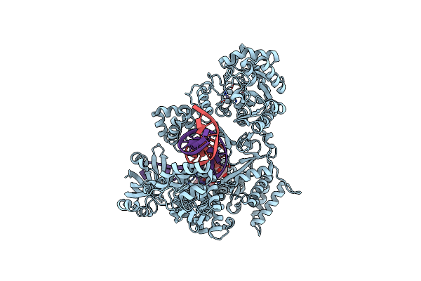 |
Organism: Mycobacterium tuberculosis, Synthetic construct
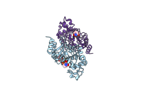
Method: ELECTRON MICROSCOPY Release Date: 2022-02-23 Classification: REPLICATION Ligands: ZN, 82W |
 |
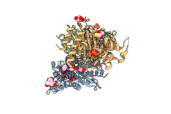
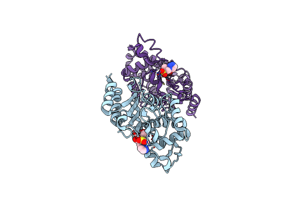
Organism: Mycobacterium tuberculosis
Method: X-RAY DIFFRACTION Resolution:1.63 Å Release Date: 2019-01-23 Classification: HYDROLASE Ligands: EDO, PG0 |
 |
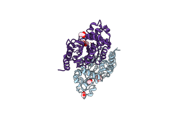
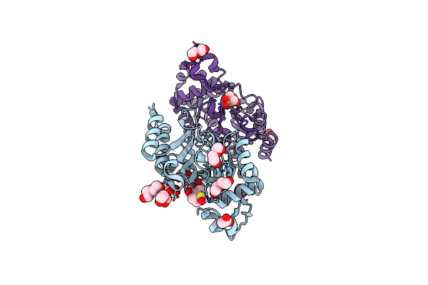
Structure Of S70A Blac From Mycobacterium Tuberculosis Obtained From Crystals Produced In The Absence Of Dtt
Organism: Mycobacterium tuberculosis
Method: X-RAY DIFFRACTION Resolution:1.19 Å Release Date: 2019-01-23 Classification: HYDROLASE Ligands: 15P, PEG, GOL |
 |
Structure Of S70A Blac From Mycobacterium Tuberculosis Obtained From Crystals Produced In The Presence Of Dtt
Organism: Mycobacterium tuberculosis
Method: X-RAY DIFFRACTION Resolution:2.54 Å Release Date: 2019-01-23 Classification: HYDROLASE Ligands: TAM, DTT, PO4, GOL |
 |
Structure Of Blac From Mycobacterium Tuberculosis Bound To The Trans-Enamine Adduct Derived From Clavulanic Acid.
Organism: Mycobacterium tuberculosis
Method: X-RAY DIFFRACTION Resolution:1.93 Å Release Date: 2019-01-23 Classification: HYDROLASE Ligands: ISS, ACT, GOL, 15P, CO2 |
 |
Structure Of Blac From Mycobacterium Tuberculosis Bound To The Propionaldehyde Ester Adduct Of Clavulanic Acid.
Organism: Mycobacterium tuberculosis
Method: X-RAY DIFFRACTION Release Date: 2019-01-23 Classification: HYDROLASE Ligands: FK2, ACT |
 |
Structure Of Blac From Mycobacterium Tuberculosis Covalently Bound To Avibactam.
Organism: Mycobacterium tuberculosis
Method: X-RAY DIFFRACTION Resolution:1.62 Å Release Date: 2019-01-23 Classification: HYDROLASE Ligands: NXL, 15P |
 |
Structure Of Blac From Mycobacterium Tuberculosis Bound To The Trans-Enamine Adduct Of Tazobactam.
Organism: Mycobacterium tuberculosis
Method: X-RAY DIFFRACTION Resolution:2.72 Å Release Date: 2019-01-23 Classification: HYDROLASE Ligands: TBE, ACT |
 |
Structure Of Blac From Mycobacterium Tuberculosis Bound To The Trans-Enamine Adduct Of Sulbactam.
Organism: Mycobacterium tuberculosis
Method: X-RAY DIFFRACTION Resolution:1.90 Å Release Date: 2019-01-23 Classification: HYDROLASE Ligands: TSL, PEG, GOL, ACT |
 |
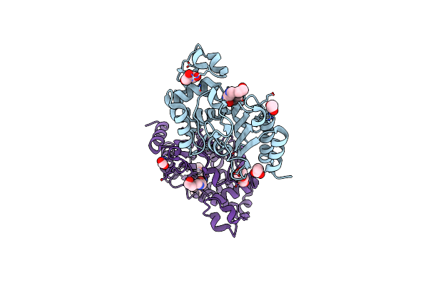
Crystal Structure Of Blac From Mycobacterium Tuberculosis Bound To Phosphate
Organism: Mycobacterium tuberculosis
Method: X-RAY DIFFRACTION Resolution:1.19 Å Release Date: 2017-11-15 Classification: HYDROLASE Ligands: PO4, ACT, ETE, AE4 |
 |
Organism: Mycobacterium tuberculosis
Method: X-RAY DIFFRACTION Resolution:2.10 Å Release Date: 2017-11-15 Classification: HYDROLASE Ligands: ACT, GOL, PGE |
 |
Organism: Streptomyces coelicolor a3(2)
Method: X-RAY DIFFRACTION Resolution:2.80 Å Release Date: 2016-12-21 Classification: ISOMERASE Ligands: PLP, CL |
 |
Alanine Racemase From Streptomyces Coelicolor A3(2) With Bound Propionate Inhibitor
Organism: Streptomyces coelicolor a3(2)
Method: X-RAY DIFFRACTION Resolution:1.51 Å Release Date: 2016-12-21 Classification: ISOMERASE Ligands: PLP, PPI, NA, NO3 |
 |
Alanine Racemase From Streptomyces Coelicolor A3(2) In Complex With D-Cycloserine
Organism: Streptomyces coelicolor a3(2)
Method: X-RAY DIFFRACTION Resolution:1.64 Å Release Date: 2016-12-21 Classification: ISOMERASE Ligands: DCS, NA, CL |