
Search Count: 466
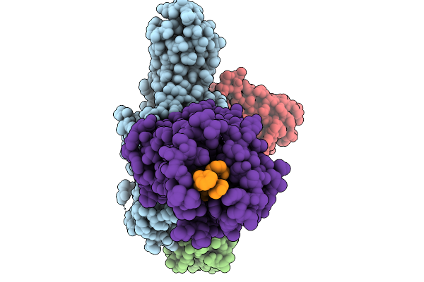 |
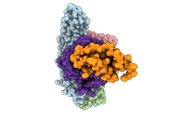
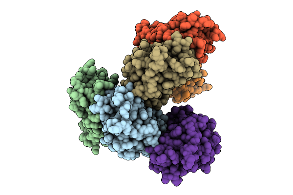
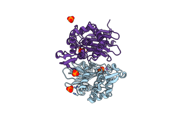
Cryo-Em Structure Of The G Protein-Coupled Receptor 1 (Gpr1) Bound To Chemerin And Beta-Arrestin 1 (Conformation 1)
Organism: Homo sapiens, Escherichia phage ecszw-2
Method: ELECTRON MICROSCOPY Resolution:3.30 Å Release Date: 2025-11-19 Classification: MEMBRANE PROTEIN/IMMUNE SYSTEM |
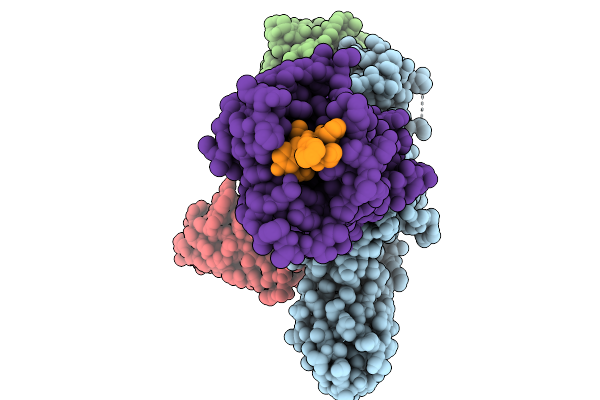 |
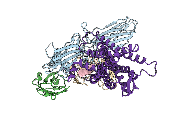
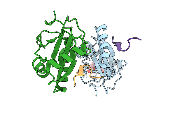
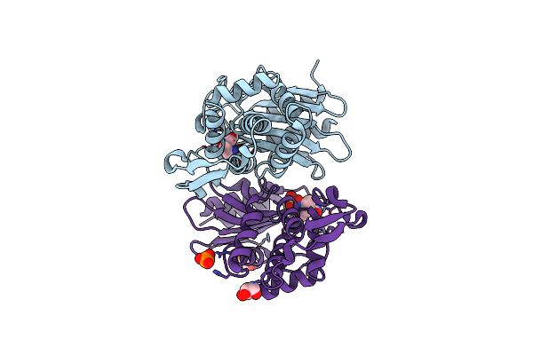
Cryo-Em Structure Of The G Protein-Coupled Receptor 1 (Gpr1) Bound To Chemerin And Beta-Arrestin 1 (Conformation 2)
Organism: Homo sapiens, Escherichia phage ecszw-2
Method: ELECTRON MICROSCOPY Resolution:3.20 Å Release Date: 2025-11-19 Classification: MEMBRANE PROTEIN/IMMUNE SYSTEM |
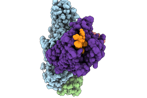 |
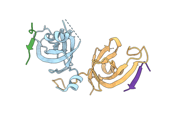
Cryo-Em Structure Of The G Protein-Coupled Receptor 1 (Gpr1) Bound To Chemerin And Beta-Arrestin 1 (Conformation 3)
Organism: Homo sapiens, Escherichia phage ecszw-2
Method: ELECTRON MICROSCOPY Resolution:3.30 Å Release Date: 2025-11-19 Classification: MEMBRANE PROTEIN/IMMUNE SYSTEM |
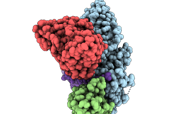 |
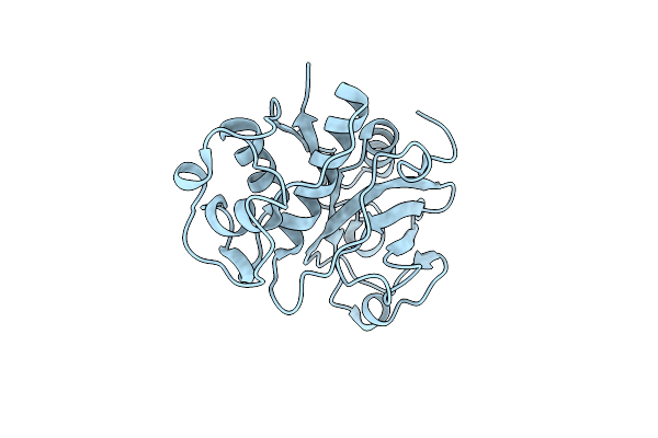
Cryo-Em Structure Of The G Protein-Coupled Receptor 1 (Gpr1) Bound To Chemerin And Beta-Arrestin 1 (Conformation 4)
Organism: Homo sapiens, Escherichia phage ecszw-2
Method: ELECTRON MICROSCOPY Resolution:3.30 Å Release Date: 2025-11-19 Classification: MEMBRANE PROTEIN/IMMUNE SYSTEM |
 |
Composite Map Of The G Protein-Coupled Receptor 1 (Gpr1) Bound To Chemerin And Beta-Arrestin 2
Organism: Homo sapiens, Escherichia phage ecszw-2
Method: ELECTRON MICROSCOPY Resolution:3.50 Å Release Date: 2025-11-19 Classification: MEMBRANE PROTEIN/IMMUNE SYSTEM Ligands: Y01 |
 |
Cryo-Em Structure Of The G Protein-Coupled Receptor 1 (Gpr1) Bound To Beta-Arrestin 1 In Ligand-Free State
Organism: Homo sapiens, Escherichia phage ecszw-2
Method: ELECTRON MICROSCOPY Release Date: 2025-11-19 Classification: MEMBRANE PROTEIN/IMMUNE SYSTEM Ligands: PAM |
 |
Organism: Sordaria araneosa
Method: X-RAY DIFFRACTION Resolution:1.76 Å Release Date: 2025-10-22 Classification: BIOSYNTHETIC PROTEIN Ligands: A1LYT, MPD |
 |
The Structure Of Sdng Covalently Binding With The Cope Rearrangement Product
Organism: Sordaria araneosa
Method: X-RAY DIFFRACTION Resolution:1.72 Å Release Date: 2025-10-22 Classification: BIOSYNTHETIC PROTEIN Ligands: MPD, A1LZG |
 |
Organism: Sordaria araneosa
Method: X-RAY DIFFRACTION Resolution:1.33 Å Release Date: 2025-10-22 Classification: BIOSYNTHETIC PROTEIN Ligands: A1LZH, A1LZK |
 |
Organism: Sordaria araneosa
Method: X-RAY DIFFRACTION Resolution:1.43 Å Release Date: 2025-10-22 Classification: BIOSYNTHETIC PROTEIN Ligands: MPD |
 |
Organism: Sordaria araneosa
Method: X-RAY DIFFRACTION Resolution:2.20 Å Release Date: 2025-10-22 Classification: BIOSYNTHETIC PROTEIN Ligands: A1LYT |
 |
Structure Of Fab Hb420 In Complex With Influenza H3N2 A/Moscow/10/1999 Neuraminidase
Organism: Influenza a virus, Homo sapiens
Method: ELECTRON MICROSCOPY Release Date: 2025-10-01 Classification: VIRAL PROTEIN/IMMUNE SYSTEM |
 |
Organism: Homo sapiens
Method: X-RAY DIFFRACTION Resolution:1.76 Å Release Date: 2025-09-10 Classification: PROTEIN BINDING Ligands: MPD |
 |
Organism: Arabidopsis thaliana
Method: X-RAY DIFFRACTION Resolution:2.45 Å Release Date: 2025-09-10 Classification: LYASE Ligands: FES |
 |
Organism: Homo sapiens
Method: X-RAY DIFFRACTION Resolution:1.61 Å Release Date: 2025-08-13 Classification: PROTEIN BINDING |
 |
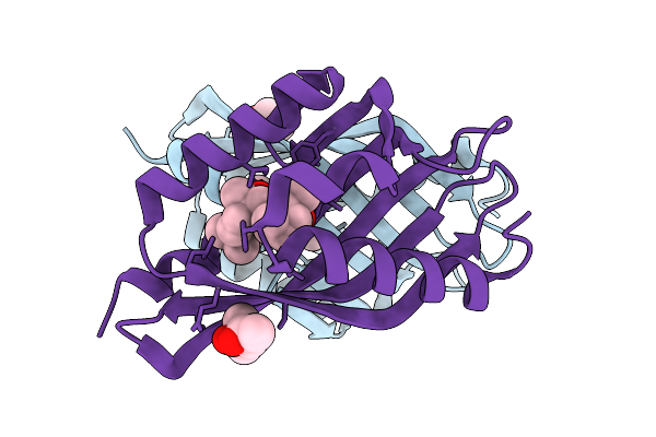
Crystal Structure Of Homolog Of Dihydroxyacid Dehydratase(Astd) From Aspergillus Terreus
Organism: Aspergillus terreus
Method: X-RAY DIFFRACTION Resolution:2.29 Å Release Date: 2025-07-09 Classification: LYASE Ligands: PEG, EDO |
 |
Organism: Carica papaya
Method: X-RAY DIFFRACTION Resolution:1.80 Å Release Date: 2025-06-18 Classification: HYDROLASE |
 |
Organism: Escherichia coli
Method: X-RAY DIFFRACTION Resolution:1.50 Å Release Date: 2025-06-18 Classification: HYDROLASE Ligands: PO4, K |
 |
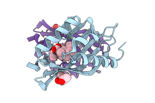
X-Ray Diffraction Structure Of Ctx-M-14 Beta-Lactamase Co-Crystallized With Avibactam
Organism: Escherichia coli
Method: X-RAY DIFFRACTION Resolution:1.60 Å Release Date: 2025-06-18 Classification: HYDROLASE Ligands: NXL, GOL, PO4 |
 |
X-Ray Diffraction Structure Of Ctx-M-14 Beta-Lactamase Soaked With Avibactam
Organism: Escherichia coli
Method: X-RAY DIFFRACTION Resolution:1.50 Å Release Date: 2025-06-18 Classification: HYDROLASE Ligands: NXL, PO4 |