
Search Count: 341
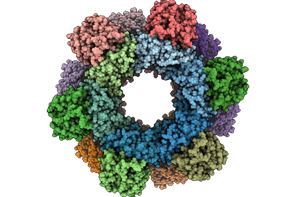 |
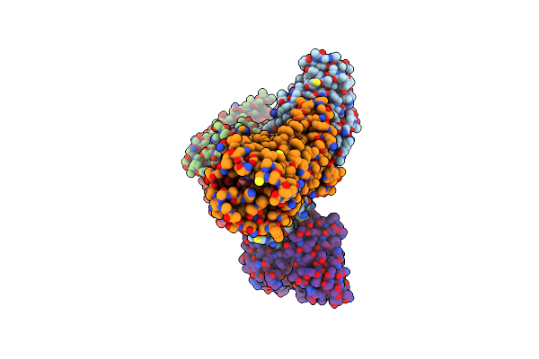
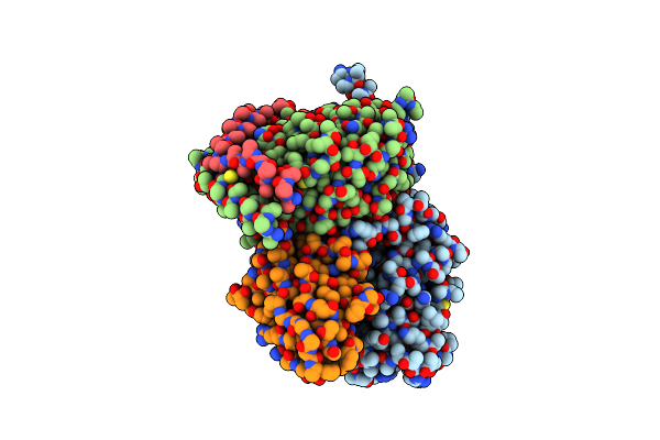
Cryo-Em Structure Of A Designed Pyridoxal Phosphate (Plp) Synthase Fused To A Designed Circumsporozoite Protein Antigen From Plasmodium Falciparum (Csp-P1-Csp And Csp-P2-Csp)
Organism: Plasmodium falciparum
Method: ELECTRON MICROSCOPY Release Date: 2025-12-31 Classification: BIOSYNTHETIC PROTEIN |
 |
Organism: Homo sapiens
Method: ELECTRON MICROSCOPY Release Date: 2025-09-10 Classification: MEMBRANE PROTEIN/IMMUNE SYSTEM Ligands: ADP, CLR |
 |
Organism: Homo sapiens, Escherichia coli
Method: ELECTRON MICROSCOPY Release Date: 2025-08-06 Classification: MEMBRANE PROTEIN/IMMUNE SYSTEM |
 |
Organism: Homo sapiens
Method: X-RAY DIFFRACTION Resolution:1.87 Å Release Date: 2025-07-30 Classification: ONCOPROTEIN Ligands: MG, A1EN3, GDP |
 |
Organism: Homo sapiens
Method: X-RAY DIFFRACTION Resolution:1.80 Å Release Date: 2025-07-30 Classification: ONCOPROTEIN Ligands: MG, A1EN3, GDP, PEG |
 |
Organism: Homo sapiens
Method: X-RAY DIFFRACTION Resolution:2.35 Å Release Date: 2025-07-30 Classification: ONCOPROTEIN Ligands: MG, A1EN3, GNP |
 |
Organism: Homo sapiens
Method: ELECTRON MICROSCOPY Release Date: 2025-06-18 Classification: HYDROLASE Ligands: NAG |
 |
Organism: Homo sapiens
Method: ELECTRON MICROSCOPY Release Date: 2025-06-18 Classification: HYDROLASE Ligands: NAG |
 |
Organism: Homo sapiens
Method: ELECTRON MICROSCOPY Release Date: 2025-06-18 Classification: HYDROLASE Ligands: NAG |
 |
Organism: Homo sapiens
Method: ELECTRON MICROSCOPY Release Date: 2025-06-18 Classification: HYDROLASE Ligands: NAG |
 |
Organism: Homo sapiens, Escherichia coli
Method: ELECTRON MICROSCOPY Resolution:2.88 Å Release Date: 2025-06-04 Classification: MEMBRANE PROTEIN/IMMUNE SYSTEM Ligands: UPG |
 |
Organism: Homo sapiens, Escherichia coli
Method: ELECTRON MICROSCOPY Release Date: 2025-06-04 Classification: MEMBRANE PROTEIN Ligands: NAI |
 |
Organism: Homo sapiens, Escherichia coli
Method: ELECTRON MICROSCOPY Resolution:2.76 Å Release Date: 2025-06-04 Classification: MEMBRANE PROTEIN/IMMUNE SYSTEM Ligands: UGA |
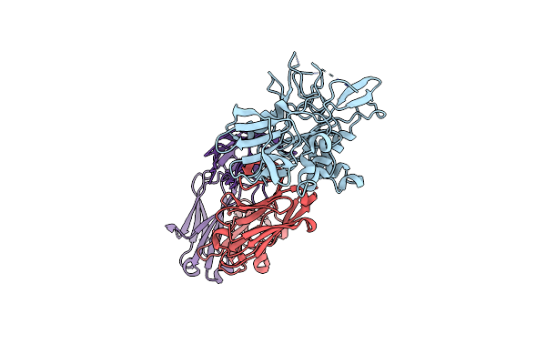 |
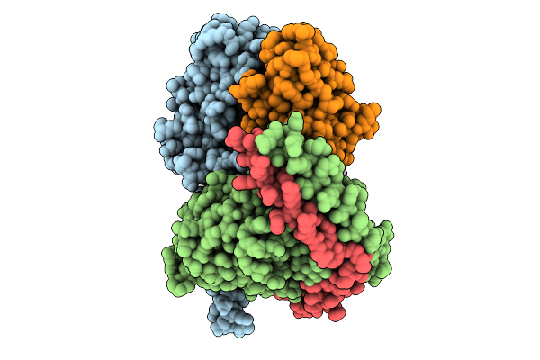
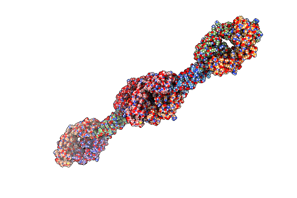
Crystal Structure Of Broadly Neutralizing Human Monoclonal Antibody 75B10 In Complex With Ama1
Organism: Plasmodium falciparum 3d7, Homo sapiens
Method: X-RAY DIFFRACTION Resolution:2.90 Å Release Date: 2025-03-12 Classification: STRUCTURAL PROTEIN |
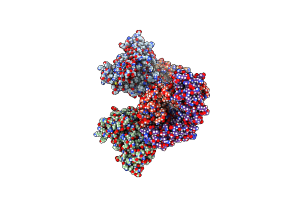 |
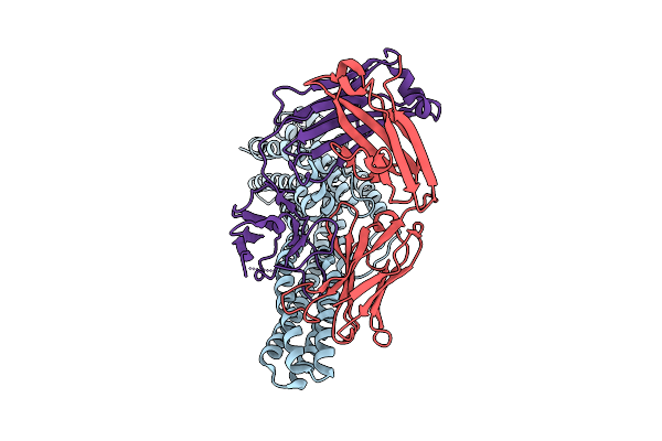
Crystal Structure Of Neutralizing Human Monoclonal Antibody 75C8 In Complex With Ama1 Di-Dii
Organism: Plasmodium falciparum 3d7, Homo sapiens
Method: X-RAY DIFFRACTION Resolution:2.80 Å Release Date: 2025-03-12 Classification: STRUCTURAL PROTEIN |
 |
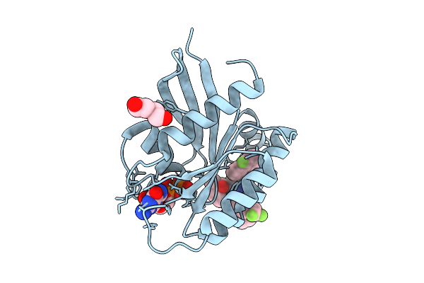
Crystal Structure Of Tet(X7) Bound To Anhydrotetracycline C10-Benzoate Ester
Organism: Pseudomonas aeruginosa
Method: X-RAY DIFFRACTION Resolution:3.30 Å Release Date: 2025-03-05 Classification: OXIDOREDUCTASE/INHIBITOR Ligands: A1BDP, FAD |
 |
Organism: Homo sapiens
Method: X-RAY DIFFRACTION Resolution:3.45 Å Release Date: 2024-10-16 Classification: RNA BINDING PROTEIN/RNA |
 |
Organism: Homo sapiens, Escherichia coli
Method: ELECTRON MICROSCOPY Resolution:3.20 Å Release Date: 2024-10-02 Classification: MEMBRANE PROTEIN Ligands: ADP, CLR |
 |
Crystal Structure Of Plasmodium Falciparum Celtos In Complex With Antibody 4H12
Organism: Plasmodium falciparum 3d7, Mus musculus
Method: X-RAY DIFFRACTION Resolution:3.52 Å Release Date: 2024-09-11 Classification: CELL INVASION |
 |
Organism: Plasmodium vivax, Mus musculus
Method: X-RAY DIFFRACTION Resolution:3.20 Å Release Date: 2024-09-11 Classification: CELL INVASION |