Search Count: 19
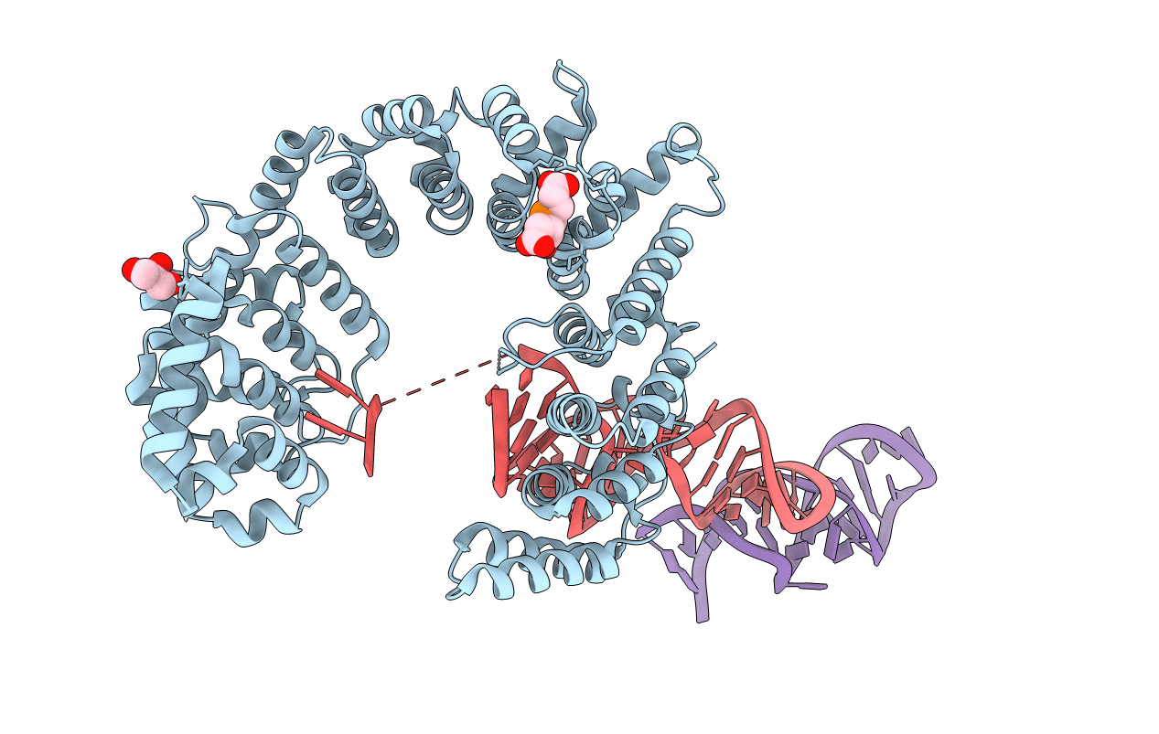 |
Organism: Saccharomyces cerevisiae (strain atcc 204508 / s288c), Saccharomyces cerevisiae s288c
Method: X-RAY DIFFRACTION Resolution:3.02 Å Release Date: 2020-05-20 Classification: RNA BINDING PROTEIN/RNA Ligands: TCE, GOL |
 |
Crystal Structure Of A Ternary Complex Of Fbf-2 With Lst-1 (Site B) And Compact Fbe Rna
Organism: Caenorhabditis elegans
Method: X-RAY DIFFRACTION Resolution:2.10 Å Release Date: 2019-08-21 Classification: RNA BINDING PROTEIN/RNA Ligands: GOL, EDO, NA |
 |
Crystal Structure Of Fbf-2 Repeat 5 Mutant (C363A, R364Y) In Complex With 8-Nt Rna
Organism: Caenorhabditis elegans
Method: X-RAY DIFFRACTION Resolution:2.85 Å Release Date: 2019-01-30 Classification: rna binding protein/rna |
 |
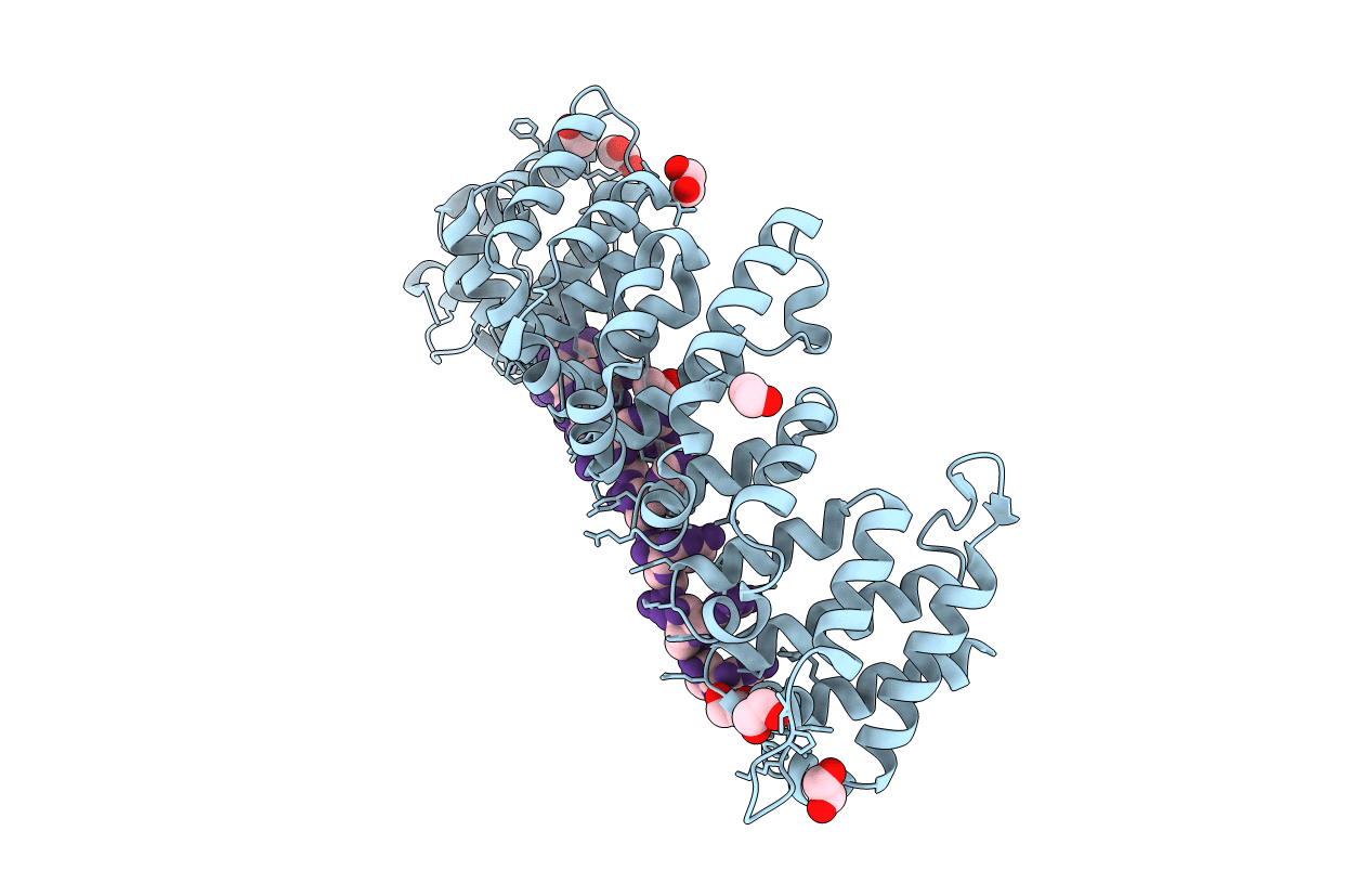
Organism: Caenorhabditis elegans
Method: X-RAY DIFFRACTION Resolution:2.55 Å Release Date: 2019-01-30 Classification: rna binding protein/rna |
 |
Crystal Structure Of Fbf-2 Repeat 5 Mutant (C363A, R364Y, Q367S) In Complex With 8-Nt Rna
Organism: Caenorhabditis elegans
Method: X-RAY DIFFRACTION Resolution:2.25 Å Release Date: 2019-01-30 Classification: rna binding protein/rna Ligands: EDO |
 |
Crystal Structure Of Fbf-2 Repeat 5 Mutant (C363S, R364Y, Q367S) In Complex With 8-Nt Rna
Organism: Caenorhabditis elegans
Method: X-RAY DIFFRACTION Resolution:2.25 Å Release Date: 2019-01-30 Classification: rna binding protein/rna Ligands: EDO |
 |
Organism: Drosophila melanogaster
Method: X-RAY DIFFRACTION Resolution:3.70 Å Release Date: 2016-08-17 Classification: RNA binding protein/RNA Ligands: ZN |
 |
Organism: Drosophila melanogaster
Method: X-RAY DIFFRACTION Resolution:4.00 Å Release Date: 2016-08-17 Classification: RNA-binding protein/RNA Ligands: ZN |
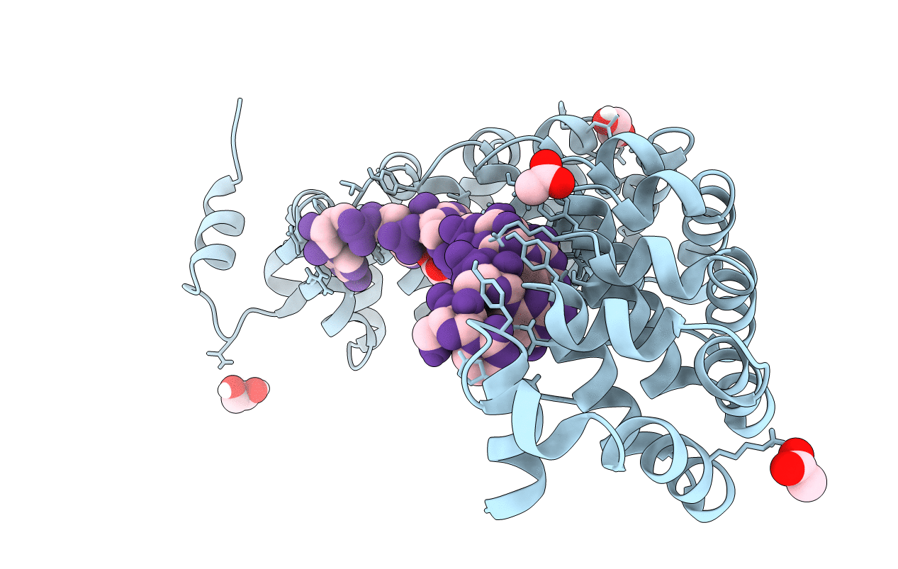 |
Crystal Structure Of The Drosophila Pumilio Rna-Binding Domain In Complex With Hunchback Rna
Organism: Drosophila melanogaster
Method: X-RAY DIFFRACTION Resolution:1.14 Å Release Date: 2016-08-17 Classification: RNA-binding protein/RNA Ligands: EDO, ACT |
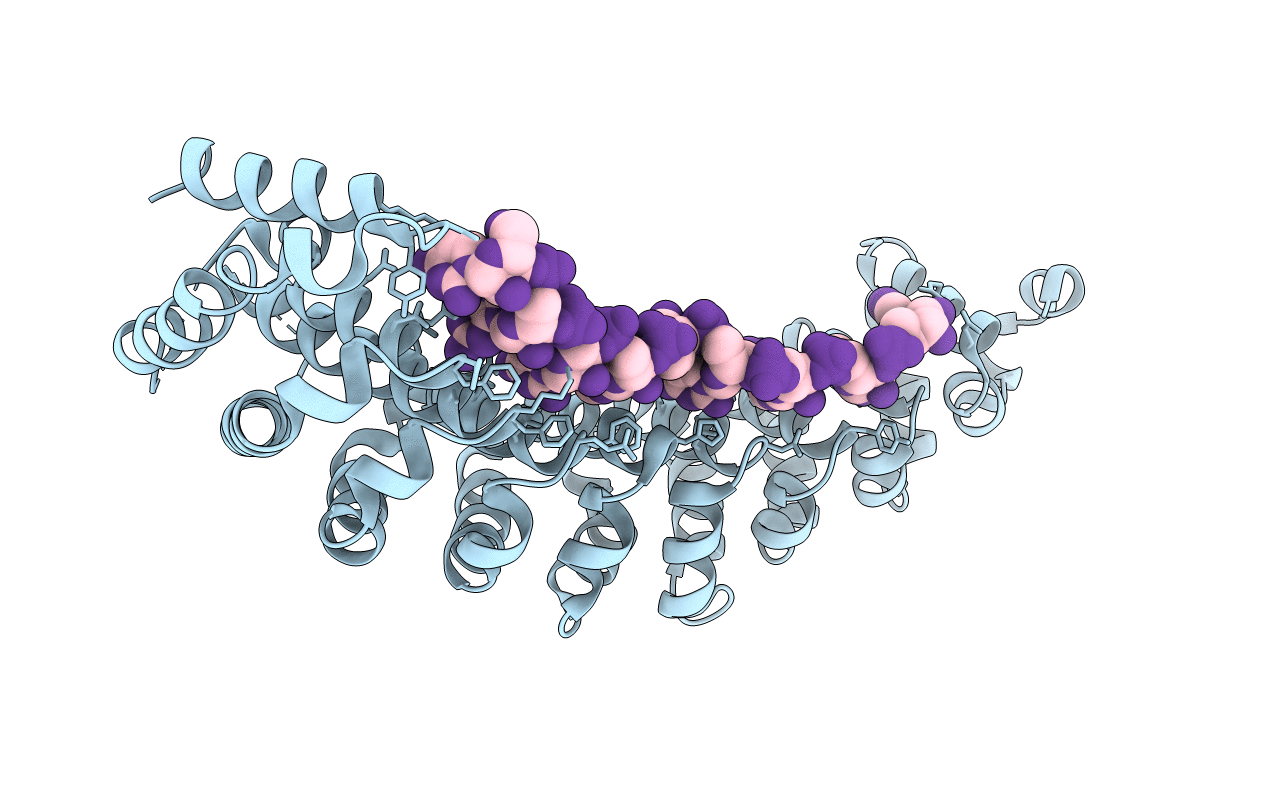 |
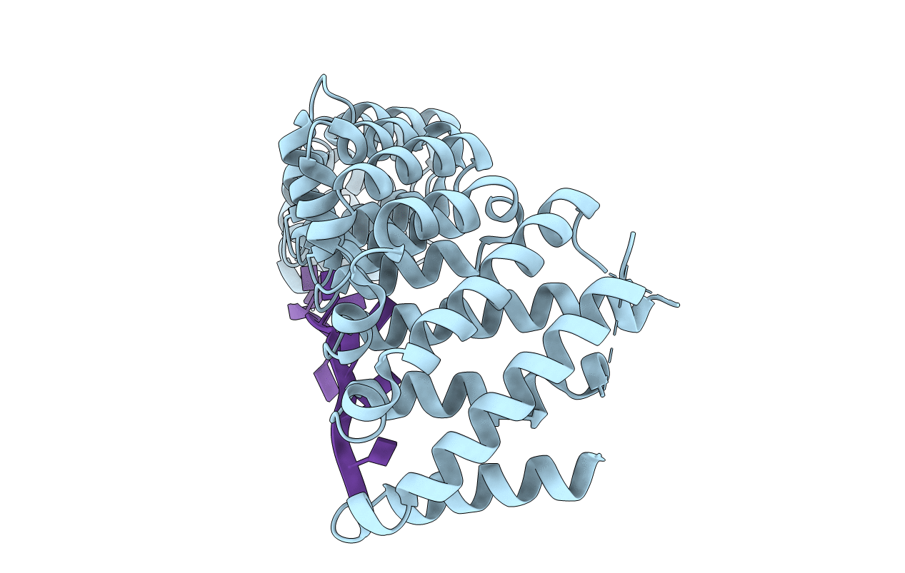
Crystal Structure Of The Rna-Binding Domain Of Yeast Puf5P Bound To Smx2 Rna
Organism: Saccharomyces cerevisiae (strain atcc 204508 / s288c), Synthetic construct
Method: X-RAY DIFFRACTION Resolution:2.71 Å Release Date: 2015-09-23 Classification: rna binding protein/rna |
 |
Crystal Structure Of The Rna-Binding Domain Of Yeast Puf5P Bound To Mfa2 Rna
Organism: Saccharomyces cerevisiae (strain atcc 204508 / s288c), Synthetic construct
Method: X-RAY DIFFRACTION Resolution:2.15 Å Release Date: 2015-09-23 Classification: rna binding protein/rna |
 |
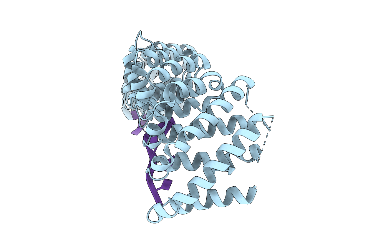
Crystal Structure Of The Rna-Binding Domain Of Yeast Puf5P Bound To Amn1 Rna
Organism: Saccharomyces cerevisiae (strain atcc 204508 / s288c), Synthetic construct
Method: X-RAY DIFFRACTION Resolution:2.80 Å Release Date: 2015-09-23 Classification: rna binding protein/rna |
 |
Crystal Structure Of The Rna-Binding Domain Of Yeast Puf5P Bound To Aat2 Rna
Organism: Saccharomyces cerevisiae (strain atcc 204508 / s288c), Synthetic construct
Method: X-RAY DIFFRACTION Resolution:2.50 Å Release Date: 2015-09-23 Classification: rna binding protein/rna |
 |
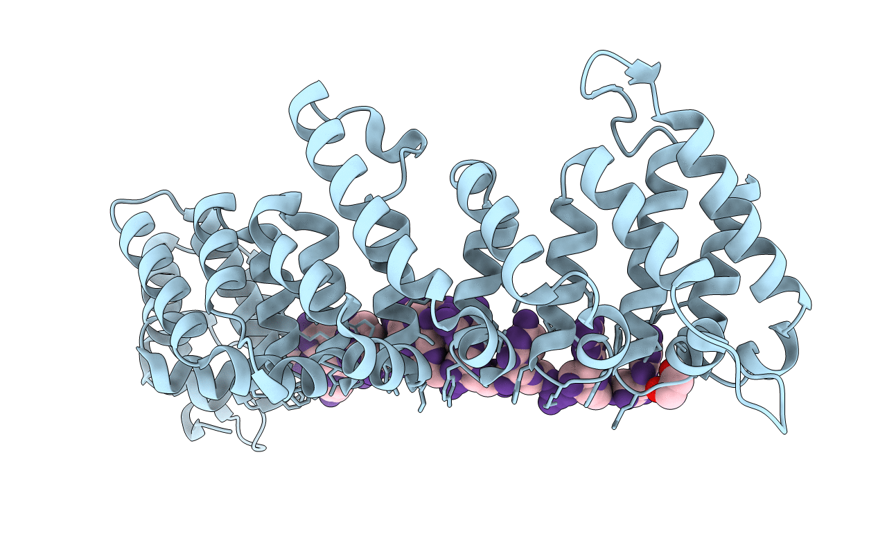
Crystal Structure Of The Rna-Binding Domain Of Yeast Puf5P Bound To Smx2 Rna
Organism: Saccharomyces cerevisiae (strain atcc 204508 / s288c), Synthetic construct
Method: X-RAY DIFFRACTION Resolution:2.35 Å Release Date: 2015-09-23 Classification: rna binding protein/rna |
 |
Organism: Caenorhabditis elegans
Method: X-RAY DIFFRACTION Resolution:2.25 Å Release Date: 2011-03-23 Classification: RNA binding protein/RNA Ligands: EDO |
 |
Organism: Caenorhabditis elegans
Method: X-RAY DIFFRACTION Resolution:2.40 Å Release Date: 2011-03-23 Classification: RNA BINDING PROTEIN/RNA |
 |
Crystal Structure Of Fbf-2 R288Y Mutant In Complex With Gld-1 Fbea A7U Mutant
Organism: Caenorhabditis elegans
Method: X-RAY DIFFRACTION Resolution:1.90 Å Release Date: 2011-03-23 Classification: RNA BINDING PROTEIN/RNA |
 |
Organism: Homo sapiens
Method: X-RAY DIFFRACTION Resolution:1.80 Å Release Date: 2001-02-05 Classification: TRANSCRIPTION/RNA |
 |
Crystal Structure Of Hud And Au-Rich Element Of The Tumor Necrosis Factor Alpha Rna
Organism: Homo sapiens
Method: X-RAY DIFFRACTION Resolution:2.30 Å Release Date: 2001-02-05 Classification: TRANSCRIPTION/RNA |

