
Search Count: 138
 |
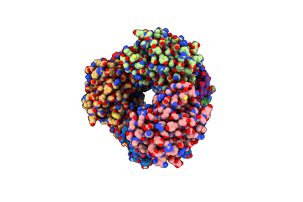
Organism: Severe acute respiratory syndrome coronavirus 2
Method: X-RAY DIFFRACTION Release Date: 2025-09-24 Classification: VIRAL PROTEIN Ligands: A1MA2, EDO |
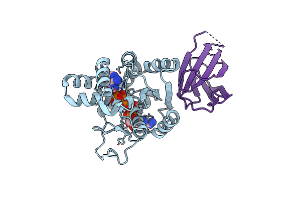 |
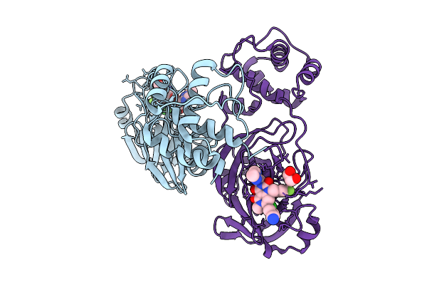
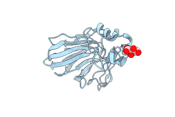
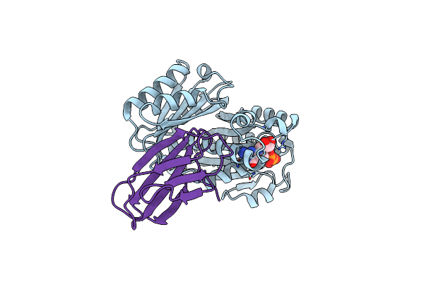
Crystal Structure Of The Monobody Cl-1 In Complex With The Escherichia Coli Adenylate Kinase
Organism: Escherichia coli (strain k12), Synthetic construct
Method: X-RAY DIFFRACTION Release Date: 2025-06-04 Classification: PROTEIN BINDING Ligands: AP5, MG |
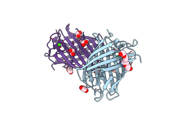 |
Crystal Structure Of Mrfp1 With A Grafted Calcium-Binding Sequence And One Bound Calcium Ion In A Calcium-Containing Solution
Organism: Discosoma, Synthetic construct
Method: X-RAY DIFFRACTION Release Date: 2025-06-04 Classification: FLUORESCENT PROTEIN Ligands: CA, EDO, PG4, PGE |
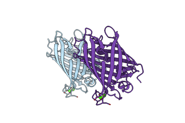 |
Crystal Structure Of Mrfp1 With A Grafted Calcium-Binding Sequence And Two Bound Calcium Ions In A Calcium-Free Solution
Organism: Discosoma, Synthetic construct
Method: X-RAY DIFFRACTION Release Date: 2025-06-04 Classification: FLUORESCENT PROTEIN Ligands: CA |
 |
Crystal Structure Of Mrfp1 With A Grafted Calcium-Binding Sequence And One Bound Calcium Ion In A Calcium-Free Solution
Organism: Discosoma, Synthetic construct
Method: X-RAY DIFFRACTION Release Date: 2025-06-04 Classification: FLUORESCENT PROTEIN Ligands: CA, PEG, EDO |
 |
Crystal Structure Of The Calcium-Free Mrfp1 With A Grafted Calcium-Binding Sequence
Organism: Discosoma, Synthetic construct
Method: X-RAY DIFFRACTION Release Date: 2025-06-04 Classification: FLUORESCENT PROTEIN |
 |
Magnetic Effects Of Thaumatin Crystals; Observation Of The Crystal Growth Of Magneto-Archimedes Levitation And Magnetic Orientation
Organism: Thaumatococcus daniellii
Method: X-RAY DIFFRACTION Resolution:0.99 Å Release Date: 2025-02-26 Classification: PLANT PROTEIN Ligands: TLA |
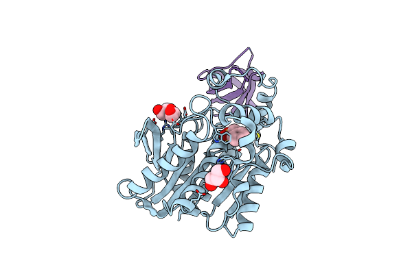 |
Crystal Structure Of The Thermotoga Maritima Cold Shock Protein Mutant Est#13 In Complex With Aacest
Organism: Alicyclobacillus acidocaldarius, Thermotoga maritima
Method: X-RAY DIFFRACTION Resolution:2.00 Å Release Date: 2025-01-15 Classification: PROTEIN BINDING Ligands: PMS, PEG, GOL |
 |
Structure Of The Sars-Cov-2 Eg.5.1 Spike Glycoprotein In Complex With Ace2 (1-Up State)
Organism: Severe acute respiratory syndrome coronavirus 2, Homo sapiens
Method: ELECTRON MICROSCOPY Release Date: 2024-05-01 Classification: VIRAL PROTEIN/PROTEIN BINDING Ligands: NAG |
 |
Organism: Homo sapiens, Severe acute respiratory syndrome coronavirus 2
Method: ELECTRON MICROSCOPY Release Date: 2024-05-01 Classification: VIRAL PROTEIN/PROTEIN BINDING Ligands: NAG |
 |
Organism: Severe acute respiratory syndrome coronavirus 2
Method: ELECTRON MICROSCOPY Release Date: 2024-04-24 Classification: VIRAL PROTEIN Ligands: NAG |
 |
Organism: Severe acute respiratory syndrome coronavirus 2
Method: ELECTRON MICROSCOPY Release Date: 2024-04-24 Classification: VIRAL PROTEIN Ligands: NAG |
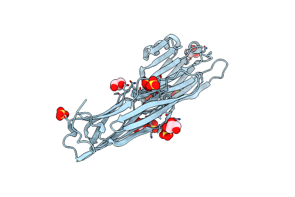 |
Crystal Structure Of The Collagen Binding Domain Of Cnm From Streptococcus Mutans
Organism: Streptococcus mutans
Method: X-RAY DIFFRACTION Resolution:1.81 Å Release Date: 2024-04-10 Classification: PROTEIN BINDING Ligands: SO4, GOL |
 |
Organism: Escherichia phage mu
Method: X-RAY DIFFRACTION Resolution:2.00 Å Release Date: 2024-02-28 Classification: VIRAL PROTEIN |
 |
Organism: Severe acute respiratory syndrome coronavirus 2
Method: ELECTRON MICROSCOPY Release Date: 2024-01-03 Classification: VIRAL PROTEIN Ligands: NAG |
 |
Organism: Severe acute respiratory syndrome coronavirus 2
Method: ELECTRON MICROSCOPY Release Date: 2024-01-03 Classification: VIRAL PROTEIN Ligands: NAG |
 |
Structure Of Sars-Cov-2 Xbb.1.5 Spike Glycoprotein In Complex With Ace2 (1-Up State)
Organism: Severe acute respiratory syndrome coronavirus 2, Homo sapiens
Method: ELECTRON MICROSCOPY Release Date: 2024-01-03 Classification: VIRAL PROTEIN/PROTEIN BINDING Ligands: NAG |
 |
Structure Of Sars-Cov-2 Xbb.1.5 Spike Glycoprotein In Complex With Ace2 (2-Up State)
Organism: Severe acute respiratory syndrome coronavirus 2, Homo sapiens
Method: ELECTRON MICROSCOPY Release Date: 2024-01-03 Classification: VIRAL PROTEIN/PROTEIN BINDING Ligands: NAG |
 |
Organism: Severe acute respiratory syndrome coronavirus 2, Homo sapiens
Method: ELECTRON MICROSCOPY Release Date: 2024-01-03 Classification: VIRAL PROTEIN/PROTEIN BINDING Ligands: NAG |
 |
Organism: Klebsiella pneumoniae, Homo sapiens
Method: ELECTRON MICROSCOPY Release Date: 2023-08-02 Classification: CELL CYCLE Ligands: GDP |