
Search Count: 28
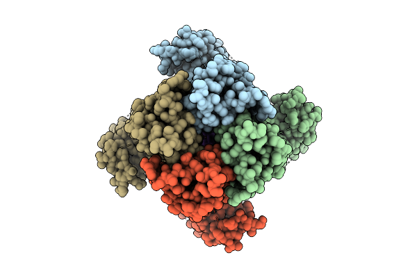 |
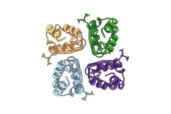
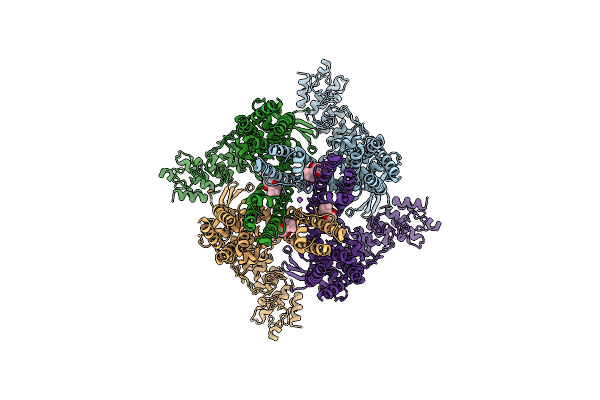
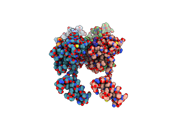
Organism: Drosophila melanogaster
Method: ELECTRON MICROSCOPY Release Date: 2025-08-06 Classification: MEMBRANE PROTEIN Ligands: POV, K |
 |
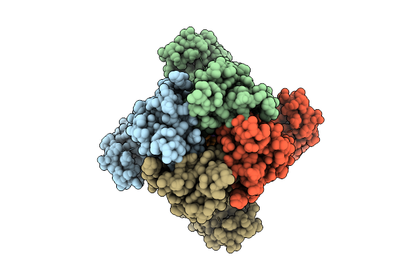
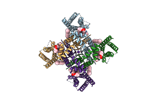
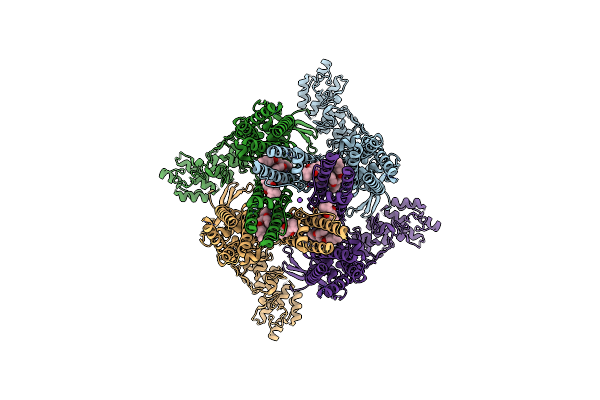
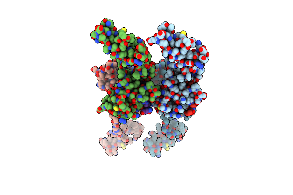
Organism: Drosophila melanogaster
Method: ELECTRON MICROSCOPY Release Date: 2025-08-06 Classification: MEMBRANE PROTEIN Ligands: POV, K |
 |
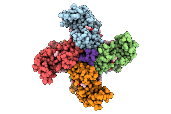
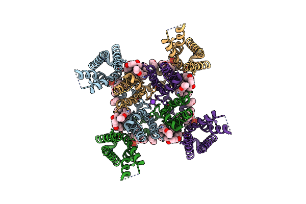
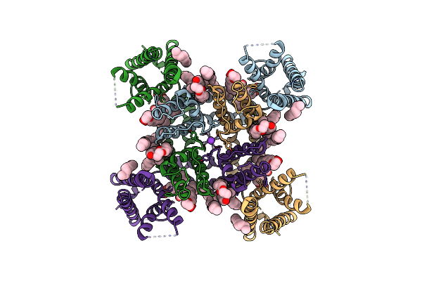
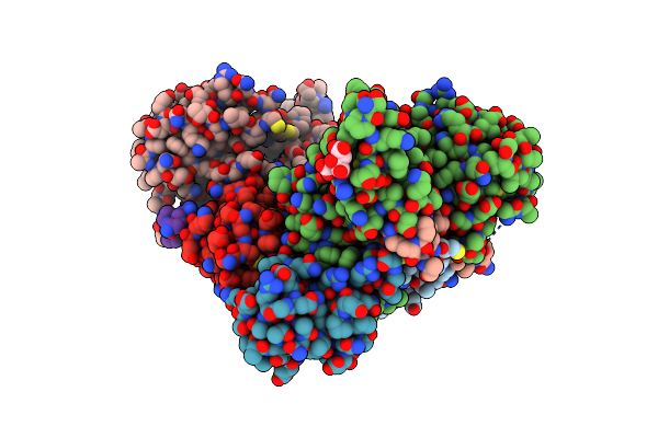
Organism: Drosophila melanogaster
Method: ELECTRON MICROSCOPY Release Date: 2025-08-06 Classification: MEMBRANE PROTEIN Ligands: POV, K |
 |
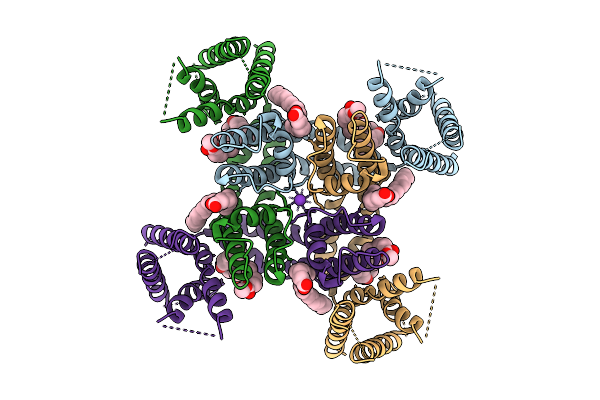
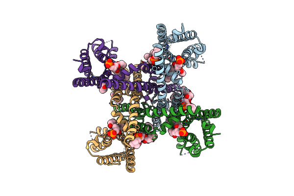
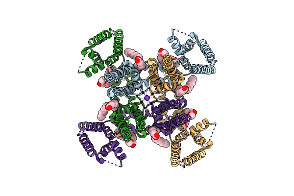
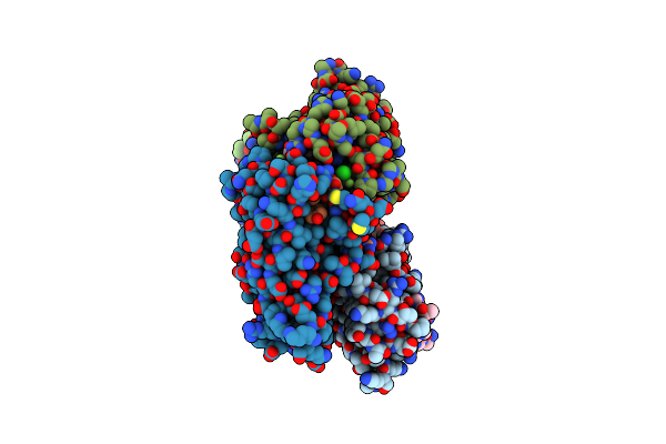
Organism: Drosophila melanogaster
Method: ELECTRON MICROSCOPY Release Date: 2025-08-06 Classification: MEMBRANE PROTEIN Ligands: POV, K |
 |
Organism: Drosophila melanogaster
Method: ELECTRON MICROSCOPY Release Date: 2025-08-06 Classification: MEMBRANE PROTEIN Ligands: POV, K |
 |
Organism: Drosophila melanogaster
Method: ELECTRON MICROSCOPY Release Date: 2025-08-06 Classification: MEMBRANE PROTEIN |
 |
Organism: Drosophila melanogaster
Method: ELECTRON MICROSCOPY Release Date: 2023-12-20 Classification: MEMBRANE PROTEIN Ligands: POV, K |
 |
Organism: Rattus norvegicus
Method: ELECTRON MICROSCOPY Release Date: 2023-09-27 Classification: MEMBRANE PROTEIN Ligands: POV, K |
 |
Organism: Rattus norvegicus
Method: ELECTRON MICROSCOPY Release Date: 2023-09-27 Classification: MEMBRANE PROTEIN Ligands: POV, K |
 |
Organism: Rattus norvegicus
Method: ELECTRON MICROSCOPY Release Date: 2023-05-31 Classification: MEMBRANE PROTEIN Ligands: P0T, NA |
 |
Organism: Rattus norvegicus
Method: ELECTRON MICROSCOPY Release Date: 2023-05-31 Classification: MEMBRANE PROTEIN Ligands: P0T, NA, POV |
 |
Organism: Drosophila melanogaster
Method: ELECTRON MICROSCOPY Release Date: 2022-03-30 Classification: MEMBRANE PROTEIN Ligands: POV, K |
 |
Organism: Drosophila melanogaster
Method: ELECTRON MICROSCOPY Release Date: 2022-03-30 Classification: MEMBRANE PROTEIN Ligands: POV, K |
 |
Organism: Bos taurus, Homo sapiens
Method: X-RAY DIFFRACTION Resolution:2.65 Å Release Date: 2021-02-17 Classification: TRANSFERASE Ligands: ZN |
 |
Organism: Bos taurus, Homo sapiens
Method: X-RAY DIFFRACTION Resolution:2.80 Å Release Date: 2021-02-17 Classification: TRANSFERASE Ligands: ZN |
 |
Crystal Structure Of Bovine Dnmt1 Rfts Domain In Complex With H3K9Me3 And Ubiquitin
Organism: Homo sapiens, Bos taurus
Method: X-RAY DIFFRACTION Resolution:3.01 Å Release Date: 2020-07-15 Classification: DNA BINDING PROTEIN Ligands: FLC, ZN |
 |
Structure Of Human Atg9A, The Only Transmembrane Protein Of The Core Autophagy Machinery
Organism: Homo sapiens
Method: ELECTRON MICROSCOPY Release Date: 2020-07-08 Classification: MEMBRANE PROTEIN Ligands: LMN |
 |
Structure Of Human Atg9A, The Only Transmembrane Protein Of The Core Autophagy Machinery
Organism: Homo sapiens
Method: ELECTRON MICROSCOPY Release Date: 2020-07-08 Classification: MEMBRANE PROTEIN Ligands: LMN |
 |
Organism: Homo sapiens, Unidentified
Method: X-RAY DIFFRACTION Resolution:3.20 Å Release Date: 2018-10-17 Classification: TRANSFERASE |
 |
Organism: Danio rerio, Homo sapiens
Method: X-RAY DIFFRACTION Resolution:1.68 Å Release Date: 2018-02-14 Classification: TRANSFERASE |