
Search Count: 75
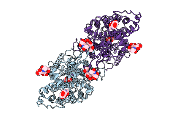 |
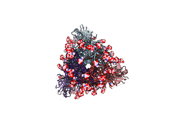
Organism: Homo sapiens
Method: ELECTRON MICROSCOPY Release Date: 2025-09-03 Classification: PROTEIN TRANSPORT Ligands: CO3, NAG, NA |
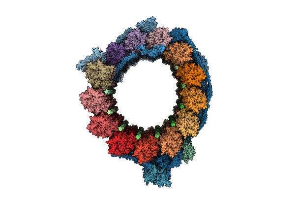 |
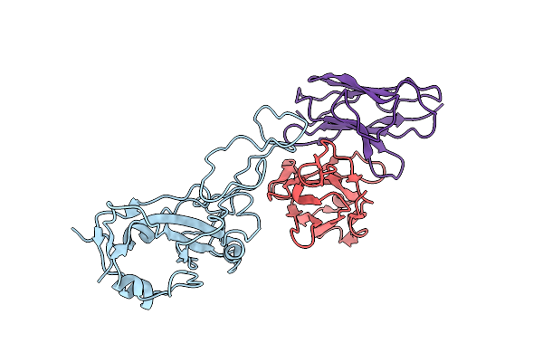
Organism: Tetrahymena thermophila cu428
Method: ELECTRON MICROSCOPY Release Date: 2025-07-30 Classification: PROTEIN BINDING Ligands: GTP, MG, GDP |
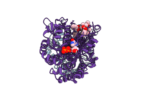 |
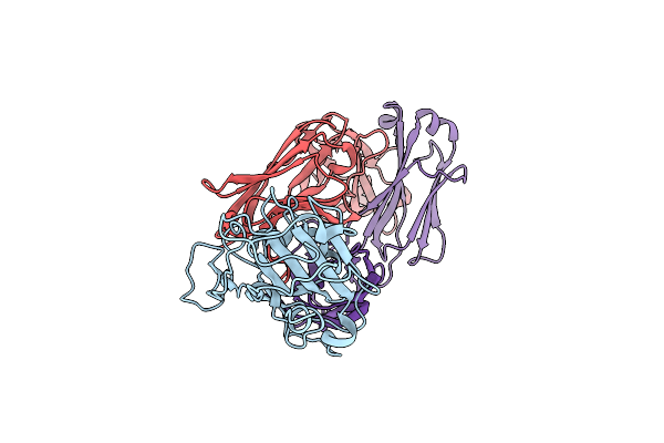
Organism: Bos taurus
Method: ELECTRON MICROSCOPY Release Date: 2025-07-30 Classification: STRUCTURAL PROTEIN Ligands: GTP, MG, GDP, TA1 |
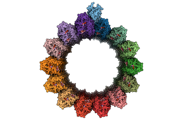 |
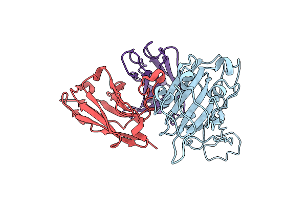
Organism: Homo sapiens, Recombinant vesicular stomatitis indiana virus rvsv-g/gfp, Bos taurus
Method: ELECTRON MICROSCOPY Release Date: 2025-07-23 Classification: PROTEIN BINDING Ligands: GTP, MG, GDP, TA1 |
 |
Organism: Plasmodium falciparum
Method: X-RAY DIFFRACTION Resolution:2.13 Å Release Date: 2025-05-21 Classification: STRUCTURAL PROTEIN |
 |
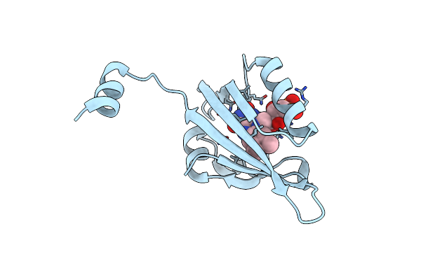
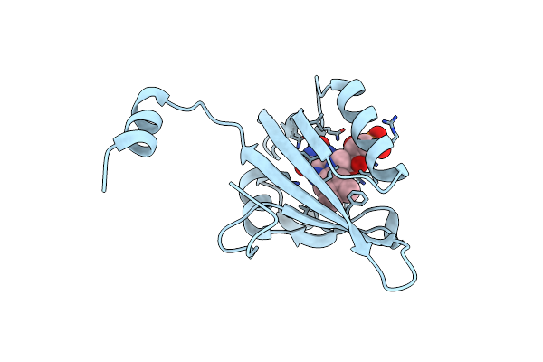
Crystal Structure Of Dh Domain Of Fyve Domain Containing Protein(Fp10) From Entamoeba Histolytica
Organism: Entamoeba histolytica hm-1:imss-a
Method: X-RAY DIFFRACTION Resolution:2.48 Å Release Date: 2025-05-21 Classification: SIGNALING PROTEIN Ligands: CIT |
 |
Crystal Structure Of The Lov1 R60K Mutant Of Klebsormidium Nitens Phototropin
Organism: Klebsormidium nitens
Method: X-RAY DIFFRACTION Resolution:1.85 Å Release Date: 2024-05-01 Classification: FLAVOPROTEIN Ligands: FMN |
 |
Crystal Structure Of Lov1 D33N Mutant Of Phototropin From Klebsormidium Nitens
Organism: Klebsormidium nitens
Method: X-RAY DIFFRACTION Resolution:2.08 Å Release Date: 2024-04-10 Classification: FLAVOPROTEIN Ligands: FMN |
 |
Crystal Structure Of The Lov1 Q122N Mutant Of Klebsormidium Nitens Phototropin
Organism: Klebsormidium nitens
Method: X-RAY DIFFRACTION Resolution:2.16 Å Release Date: 2024-03-06 Classification: FLAVOPROTEIN Ligands: FMN |
 |
Organism: Klebsormidium nitens
Method: X-RAY DIFFRACTION Resolution:1.86 Å Release Date: 2024-01-24 Classification: FLAVOPROTEIN Ligands: FMN |
 |
Organism: Severe acute respiratory syndrome coronavirus 2
Method: ELECTRON MICROSCOPY Release Date: 2023-06-07 Classification: VIRAL PROTEIN Ligands: NAG |
 |
Organism: Severe acute respiratory syndrome coronavirus 2
Method: ELECTRON MICROSCOPY Release Date: 2023-06-07 Classification: VIRAL PROTEIN Ligands: NAG |
 |
Organism: Severe acute respiratory syndrome coronavirus 2
Method: ELECTRON MICROSCOPY Release Date: 2023-06-07 Classification: VIRAL PROTEIN Ligands: NAG |
 |
Organism: Severe acute respiratory syndrome coronavirus 2, Vicugna pacos
Method: X-RAY DIFFRACTION Resolution:3.00 Å Release Date: 2022-06-29 Classification: VIRAL PROTEIN Ligands: GOL, PO4 |
 |
Organism: Severe acute respiratory syndrome coronavirus 2, Vicugna pacos
Method: X-RAY DIFFRACTION Resolution:1.75 Å Release Date: 2022-05-25 Classification: VIRAL PROTEIN Ligands: GOL, ACT |
 |
Organism: Severe acute respiratory syndrome coronavirus 2, Homo sapiens
Method: ELECTRON MICROSCOPY Release Date: 2022-04-27 Classification: VIRAL PROTEIN/Immune System |
 |
Organism: Severe acute respiratory syndrome coronavirus 2, Homo sapiens
Method: ELECTRON MICROSCOPY Release Date: 2022-04-27 Classification: VIRAL PROTEIN/Immune System |
 |
Organism: Severe acute respiratory syndrome coronavirus 2, Homo sapiens
Method: ELECTRON MICROSCOPY Release Date: 2022-04-27 Classification: VIRAL PROTEIN/Immune System |
 |
Organism: Severe acute respiratory syndrome coronavirus 2
Method: ELECTRON MICROSCOPY Release Date: 2022-02-16 Classification: VIRAL PROTEIN Ligands: NAG |
 |
Organism: Severe acute respiratory syndrome coronavirus 2
Method: ELECTRON MICROSCOPY Release Date: 2022-02-16 Classification: VIRAL PROTEIN Ligands: NAG |