
Search Count: 34
 |
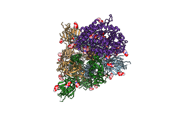
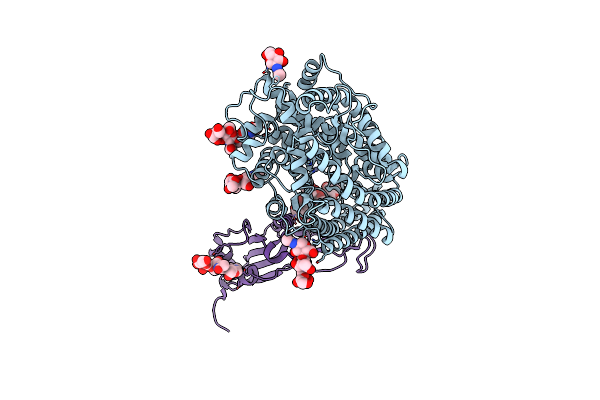
Cryo-Em Structure Of Tmprss2 In Complex With Fab Fragments Of 752 Mab And 2228 Mab
Organism: Homo sapiens, Mus musculus
Method: ELECTRON MICROSCOPY Release Date: 2025-10-15 Classification: MEMBRANE PROTEIN |
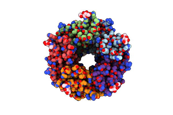 |
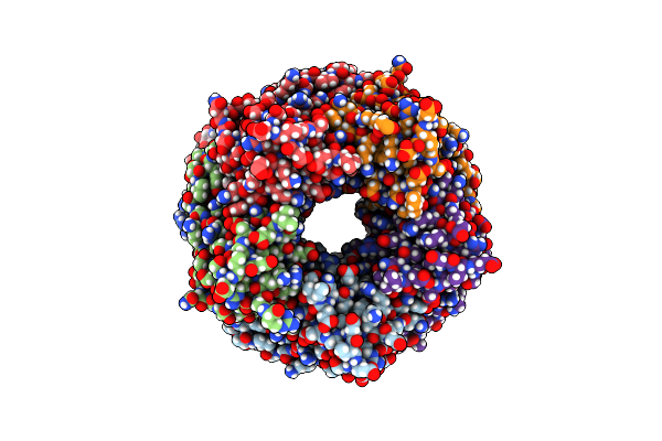
Crystal Structure Of Lymnaea Stagnalis Acetylcholine-Binding Protein Q55R Mutant Complexed With Dinotefuran
Organism: Lymnaea stagnalis
Method: X-RAY DIFFRACTION Resolution:2.63 Å Release Date: 2024-12-18 Classification: SIGNALING PROTEIN Ligands: A1LW0, NAG |
 |
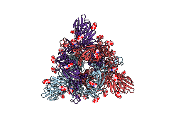
Structure Of The Sars-Cov-2 Eg.5.1 Spike Glycoprotein In Complex With Ace2 (1-Up State)
Organism: Severe acute respiratory syndrome coronavirus 2, Homo sapiens
Method: ELECTRON MICROSCOPY Release Date: 2024-05-01 Classification: VIRAL PROTEIN/PROTEIN BINDING Ligands: NAG |
 |
Organism: Homo sapiens, Severe acute respiratory syndrome coronavirus 2
Method: ELECTRON MICROSCOPY Release Date: 2024-05-01 Classification: VIRAL PROTEIN/PROTEIN BINDING Ligands: NAG |
 |
Organism: Severe acute respiratory syndrome coronavirus 2
Method: ELECTRON MICROSCOPY Release Date: 2024-04-24 Classification: VIRAL PROTEIN Ligands: NAG |
 |
Organism: Severe acute respiratory syndrome coronavirus 2
Method: ELECTRON MICROSCOPY Release Date: 2024-04-24 Classification: VIRAL PROTEIN Ligands: NAG |
 |
Organism: Severe acute respiratory syndrome coronavirus 2
Method: ELECTRON MICROSCOPY Release Date: 2024-01-03 Classification: VIRAL PROTEIN Ligands: NAG |
 |
Organism: Severe acute respiratory syndrome coronavirus 2
Method: ELECTRON MICROSCOPY Release Date: 2024-01-03 Classification: VIRAL PROTEIN Ligands: NAG |
 |
Structure Of Sars-Cov-2 Xbb.1.5 Spike Glycoprotein In Complex With Ace2 (1-Up State)
Organism: Severe acute respiratory syndrome coronavirus 2, Homo sapiens
Method: ELECTRON MICROSCOPY Release Date: 2024-01-03 Classification: VIRAL PROTEIN/PROTEIN BINDING Ligands: NAG |
 |
Structure Of Sars-Cov-2 Xbb.1.5 Spike Glycoprotein In Complex With Ace2 (2-Up State)
Organism: Severe acute respiratory syndrome coronavirus 2, Homo sapiens
Method: ELECTRON MICROSCOPY Release Date: 2024-01-03 Classification: VIRAL PROTEIN/PROTEIN BINDING Ligands: NAG |
 |
Organism: Severe acute respiratory syndrome coronavirus 2, Homo sapiens
Method: ELECTRON MICROSCOPY Release Date: 2024-01-03 Classification: VIRAL PROTEIN/PROTEIN BINDING Ligands: NAG |
 |
Organism: Severe acute respiratory syndrome coronavirus 2
Method: ELECTRON MICROSCOPY Release Date: 2023-05-24 Classification: VIRAL PROTEIN Ligands: NAG |
 |
Organism: Severe acute respiratory syndrome coronavirus 2
Method: ELECTRON MICROSCOPY Release Date: 2023-05-24 Classification: VIRAL PROTEIN Ligands: NAG |
 |
Structure Of Sars-Cov-2 Xbb.1 Spike Glycoprotein In Complex With Ace2 (1-Up State)
Organism: Severe acute respiratory syndrome coronavirus 2, Homo sapiens
Method: ELECTRON MICROSCOPY Release Date: 2023-05-24 Classification: VIRAL PROTEIN/PROTEIN BINDING Ligands: NAG |
 |
Organism: Homo sapiens, Severe acute respiratory syndrome coronavirus 2
Method: ELECTRON MICROSCOPY Release Date: 2023-05-24 Classification: VIRAL PROTEIN/PROTEIN BINDING Ligands: NAG |
 |
Crystal Structure Of The Receptor Binding Domain Of Sars-Cov-2 Omicron Bq.1.1 Variant Spike Protein In Complex With Its Receptor Ace2
Organism: Homo sapiens, Severe acute respiratory syndrome coronavirus 2
Method: X-RAY DIFFRACTION Resolution:2.78 Å Release Date: 2023-05-17 Classification: VIRAL PROTEIN Ligands: ZN, NAG |
 |
Organism: Severe acute respiratory syndrome coronavirus 2
Method: ELECTRON MICROSCOPY Release Date: 2022-10-26 Classification: VIRAL PROTEIN Ligands: NAG |
 |
Crystal Structure Of Lymnaea Stagnalis Acetylcholine Binding Protein (Achbp) Complexed With Paraherquamide A
Organism: Lymnaea stagnalis
Method: X-RAY DIFFRACTION Resolution:2.20 Å Release Date: 2021-11-24 Classification: SIGNALING PROTEIN Ligands: H8U, NAG |
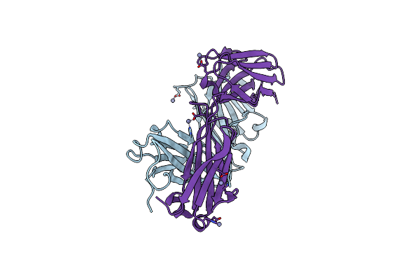 |
Crystal Structure Of The Anti-Human Prostaglandin E Receptor Ep4 Antibody Fab Fragment
Organism: Mus musculus
Method: X-RAY DIFFRACTION Resolution:1.85 Å Release Date: 2018-12-05 Classification: IMMUNE SYSTEM Ligands: ZN |
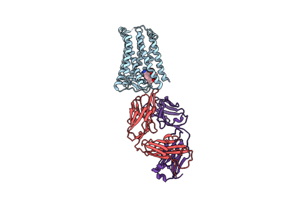 |
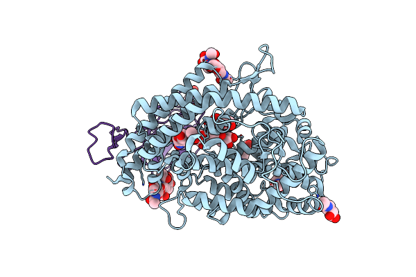
Crystal Structure Of The Human Prostaglandin E Receptor Ep4 In Complex With Fab And An Antagonist Br-Derivative
Organism: Homo sapiens, Mus musculus
Method: X-RAY DIFFRACTION Resolution:4.20 Å Release Date: 2018-12-05 Classification: SIGNALING PROTEIN/IMMUNE SYSTEM Ligands: 8VL |