
Search Count: 34
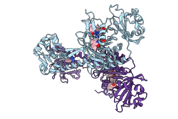 |
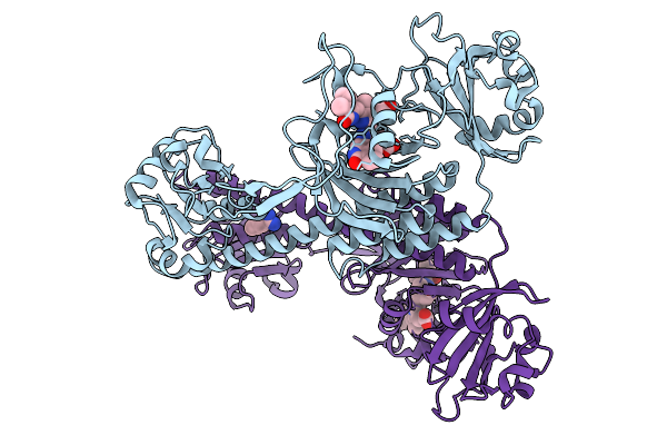
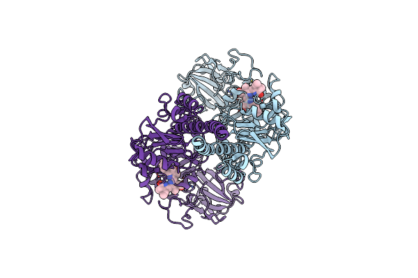
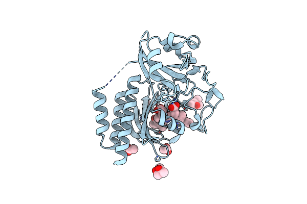
Photoactivation In Bacteriophytochromes, Reference (Dark) Structure For The 3 Ps Time Point
Organism: Stigmatella aurantiaca
Method: X-RAY DIFFRACTION Release Date: 2025-10-08 Classification: SIGNALING PROTEIN Ligands: 3Q8, BEN |
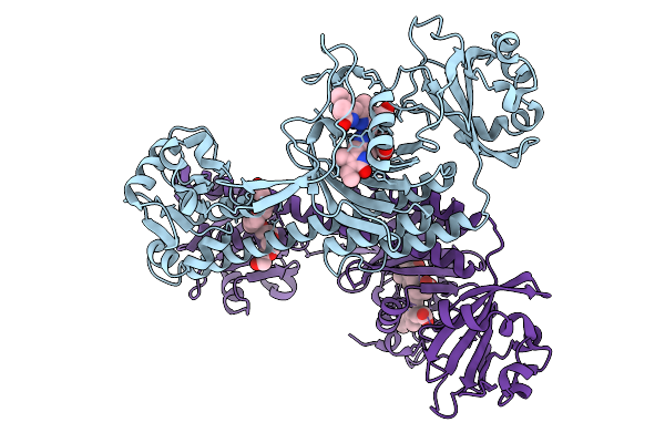 |
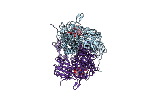
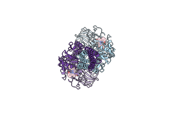
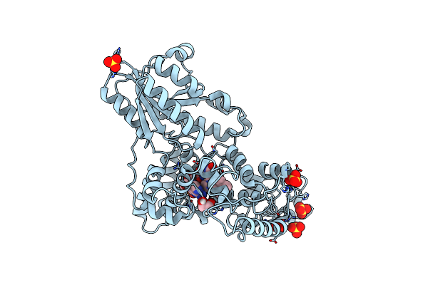
Photoactivation In Bacteriophytochrome, High Resolution Cryo Structure In The Dark.
Organism: Stigmatella aurantiaca
Method: X-RAY DIFFRACTION Release Date: 2025-10-08 Classification: SIGNALING PROTEIN Ligands: EL5, P33 |
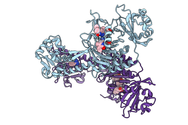 |
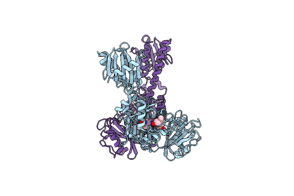
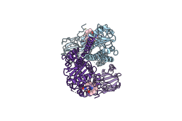
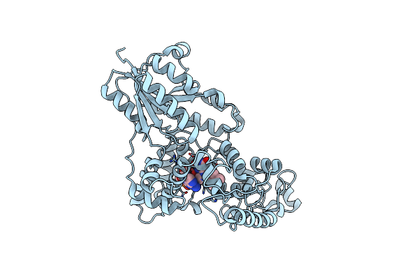
Photoactivation In Bacteriophytochromes, Reference (Dark) Structure For The 100 Ps Time Point
Organism: Stigmatella aurantiaca
Method: X-RAY DIFFRACTION Release Date: 2025-10-08 Classification: SIGNALING PROTEIN Ligands: EL5, BEN |
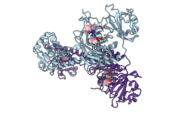 |
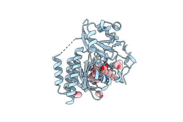
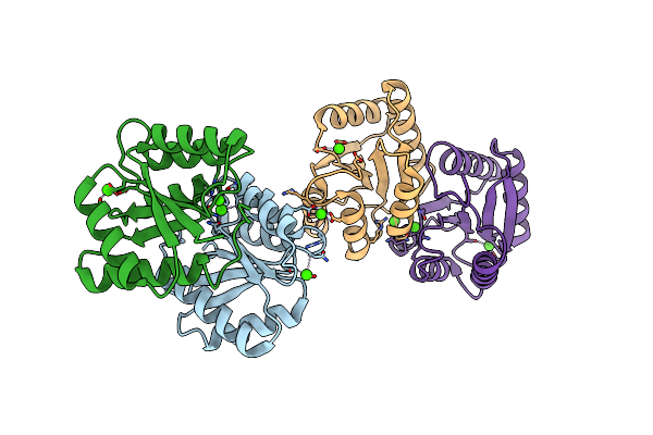
Organism: Stigmatella aurantiaca
Method: X-RAY DIFFRACTION Release Date: 2025-10-08 Classification: SIGNALING PROTEIN Ligands: BLA, BEN |
 |
Organism: Stigmatella aurantiaca
Method: X-RAY DIFFRACTION Release Date: 2025-10-08 Classification: SIGNALING PROTEIN Ligands: BLA, BEN |
 |
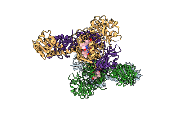
Cryo-Em Structure Of The Full-Length Pseudomonas Aeruginosa Bacteriophytochrome In Its Pr State
Organism: Pseudomonas aeruginosa
Method: ELECTRON MICROSCOPY Release Date: 2024-09-04 Classification: CYTOSOLIC PROTEIN Ligands: LBV |
 |
Cryo-Em Structure Of The Full-Length Pseudomonas Aeruginosa Bacteriophytochrome In Its Pfr State
Organism: Pseudomonas aeruginosa
Method: ELECTRON MICROSCOPY Release Date: 2024-09-04 Classification: CYTOSOLIC PROTEIN Ligands: LBV |
 |
Organism: Deinococcus radiodurans r1
Method: X-RAY DIFFRACTION Resolution:2.30 Å Release Date: 2023-06-28 Classification: TRANSFERASE Ligands: LBV |
 |
Organism: Deinococcus radiodurans r1
Method: ELECTRON MICROSCOPY Release Date: 2022-12-21 Classification: TRANSFERASE Ligands: LBV |
 |
Organism: Deinococcus radiodurans r1
Method: ELECTRON MICROSCOPY Release Date: 2022-12-21 Classification: TRANSFERASE Ligands: LBV |
 |
Organism: Deinococcus radiodurans r1
Method: ELECTRON MICROSCOPY Release Date: 2022-12-21 Classification: TRANSFERASE Ligands: BLA |
 |
Organism: Deinococcus radiodurans r1
Method: X-RAY DIFFRACTION Resolution:1.88 Å Release Date: 2022-08-10 Classification: TRANSFERASE Ligands: ACT, MPD, LBV |
 |
Organism: Deinococcus radiodurans r1
Method: X-RAY DIFFRACTION Resolution:1.48 Å Release Date: 2022-08-10 Classification: TRANSFERASE Ligands: ACT, MPD, LBV |
 |
X-Ray Crystallographic Structure Of (6-4)Photolyase From Drosophila Melanogaster At Cryogenic Temperature
Organism: Drosophila melanogaster
Method: X-RAY DIFFRACTION Resolution:1.79 Å Release Date: 2021-08-18 Classification: LYASE Ligands: GOL, FAD, SO4 |
 |
X-Ray Crystallographic Structure Of (6-4)Photolyase From Drosophila Melanogaster At Room Temperature
Organism: Drosophila melanogaster
Method: X-RAY DIFFRACTION Resolution:2.27 Å Release Date: 2021-08-18 Classification: LYASE Ligands: FAD |
 |
Organism: Deinococcus radiodurans r1
Method: X-RAY DIFFRACTION Resolution:2.10 Å Release Date: 2021-06-30 Classification: SIGNALING PROTEIN Ligands: CA |
 |
Organism: Deinococcus radiodurans
Method: X-RAY DIFFRACTION Resolution:2.07 Å Release Date: 2020-04-08 Classification: TRANSFERASE Ligands: LBV |
 |
Pas-Gaf Fragment From Deinococcus Radiodurans Phytochrome 1Ps After Photoexcitation
Organism: Deinococcus radiodurans
Method: X-RAY DIFFRACTION Resolution:2.21 Å Release Date: 2020-04-08 Classification: TRANSFERASE Ligands: LBV |
 |
Structure Of The Chromophore Binding Domain Of Stigmatella Aurantiaca Phytochrome P1, Wild-Type
Organism: Stigmatella aurantiaca dw4/3-1
Method: X-RAY DIFFRACTION Resolution:1.85 Å Release Date: 2018-09-19 Classification: SIGNALING PROTEIN Ligands: BLR |
 |
The Structure Of The Stigmatella Aurantiaca Phytochrome Chromophore Binding Domain T289H Mutant
Organism: Stigmatella aurantiaca dw4/3-1
Method: X-RAY DIFFRACTION Resolution:1.92 Å Release Date: 2018-09-19 Classification: SIGNALING PROTEIN Ligands: BLR |