Search Count: 32
 |
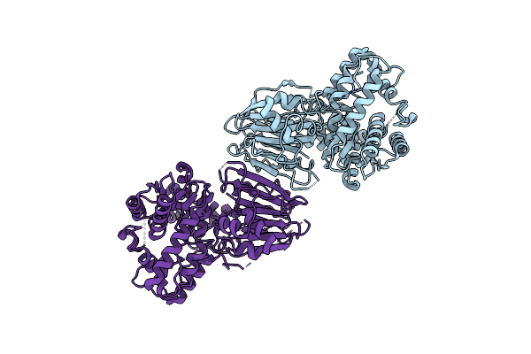
Organism: Homo sapiens
Method: ELECTRON MICROSCOPY Release Date: 2025-01-08 Classification: BIOSYNTHETIC PROTEIN |
 |
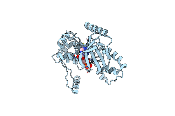
Organism: Homo sapiens
Method: ELECTRON MICROSCOPY Release Date: 2024-05-22 Classification: BIOSYNTHETIC PROTEIN |
 |
Organism: Homo sapiens
Method: X-RAY DIFFRACTION Resolution:2.01 Å Release Date: 2022-06-22 Classification: SIGNALING PROTEIN Ligands: ANP |
 |
Structure Of Rna-Dependent Rna Polymerase 2 (Rdr2) From Arabidopsis Thaliana
Organism: Arabidopsis thaliana
Method: ELECTRON MICROSCOPY Release Date: 2021-12-08 Classification: TRANSCRIPTION Ligands: MG |
 |
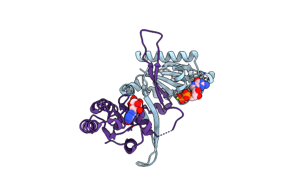
Organism: Arabidopsis thaliana
Method: ELECTRON MICROSCOPY Release Date: 2021-12-08 Classification: TRANSCRIPTION Ligands: MG |
 |
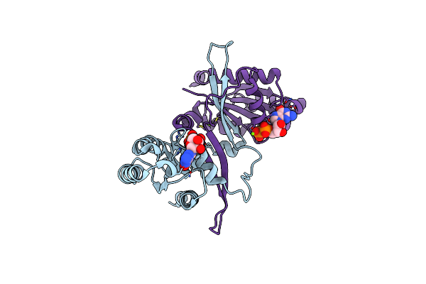
1.9A Crystal Structure Of The Gtpase Domain Of Parkinson'S Disease-Associated Protein Lrrk2 Carrying R1398H
Organism: Homo sapiens
Method: X-RAY DIFFRACTION Resolution:1.97 Å Release Date: 2021-06-09 Classification: HYDROLASE Ligands: MG, GDP |
 |
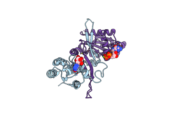
Organism: Homo sapiens
Method: X-RAY DIFFRACTION Resolution:1.95 Å Release Date: 2020-10-14 Classification: HYDROLASE Ligands: MG, GDP |
 |
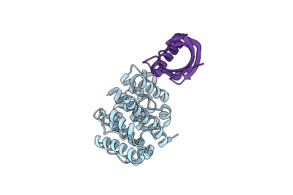
Organism: Homo sapiens
Method: X-RAY DIFFRACTION Resolution:1.60 Å Release Date: 2020-10-14 Classification: HYDROLASE Ligands: MG, GDP |
 |
Organism: Homo sapiens, Synthetic construct
Method: X-RAY DIFFRACTION Resolution:1.20 Å Release Date: 2020-10-07 Classification: CONTRACTILE PROTEIN |
 |
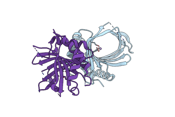
Crystal Structure Of Cet1 From Trypanosoma Cruzi In Complex With Tripolyphosphate, Manganese And Iodide Ions.
Organism: Trypanosoma cruzi strain cl brener
Method: X-RAY DIFFRACTION Resolution:2.20 Å Release Date: 2020-06-03 Classification: HYDROLASE Ligands: MN, 3PO, IOD |
 |
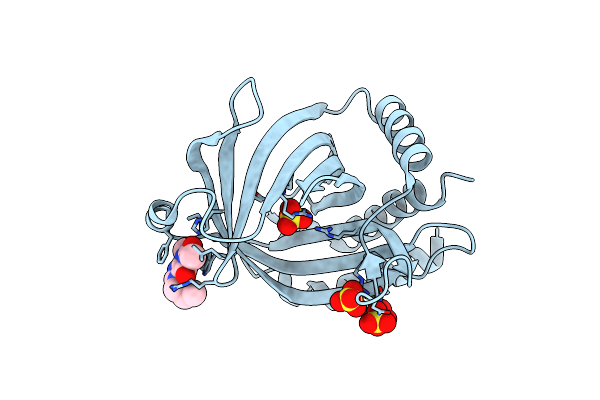
Crystal Structure Of Cet1 From Trypanosoma Cruzi In Complex With Manganese Ion.
Organism: Trypanosoma cruzi strain cl brener
Method: X-RAY DIFFRACTION Resolution:2.60 Å Release Date: 2020-06-03 Classification: HYDROLASE Ligands: MN |
 |
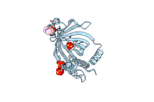
Crystal Structure Of Cet1 From Trypanosoma Cruzi In Complex With #951 Ligand
Organism: Trypanosoma cruzi strain cl brener
Method: X-RAY DIFFRACTION Resolution:2.39 Å Release Date: 2020-06-03 Classification: HYDROLASE Ligands: E7R, SO4 |
 |
Crystal Structure Of Cet1 From Trypanosoma Cruzi In Complex With #466 Ligand.
Organism: Trypanosoma cruzi strain cl brener
Method: X-RAY DIFFRACTION Resolution:2.51 Å Release Date: 2020-06-03 Classification: HYDROLASE Ligands: JJY, SO4 |
 |
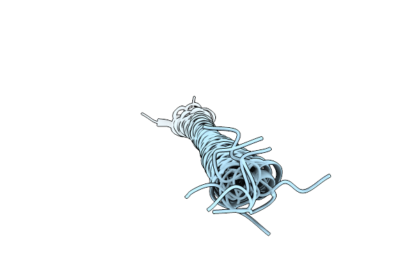
Human Asparagine Synthetase (Asns) In Complex With 6-Diazo-5-Oxo-L-Norleucine (Don) At 1.85 A Resolution
Organism: Homo sapiens
Method: X-RAY DIFFRACTION Resolution:1.85 Å Release Date: 2019-09-18 Classification: BIOSYNTHETIC PROTEIN Ligands: ONL, EDO, EPE, CL |
 |
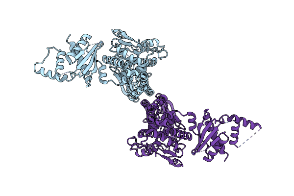
Remarkable Rigidity Of The Single Alpha-Helical Domain Of Myosin-Vi Revealed By Nmr Spectroscopy
Organism: Meriones unguiculatus
Method: SOLUTION NMR Release Date: 2019-06-12 Classification: MOTOR PROTEIN |
 |
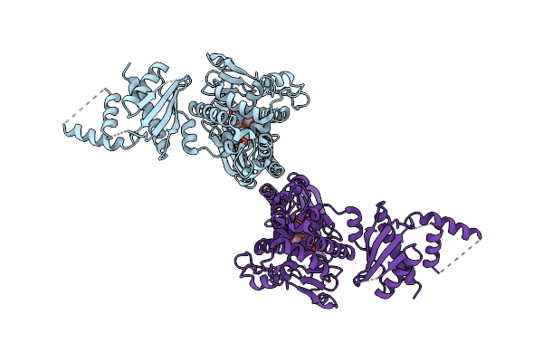
Organism: Homo sapiens
Method: X-RAY DIFFRACTION Resolution:2.80 Å Release Date: 2017-05-31 Classification: TRANSFERASE Ligands: CL |
 |
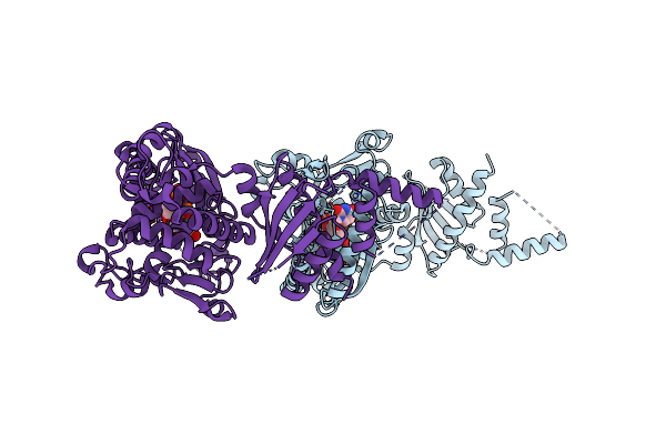
Organism: Homo sapiens
Method: X-RAY DIFFRACTION Resolution:2.95 Å Release Date: 2017-05-31 Classification: TRANSFERASE Ligands: UTP, BA, CL |
 |
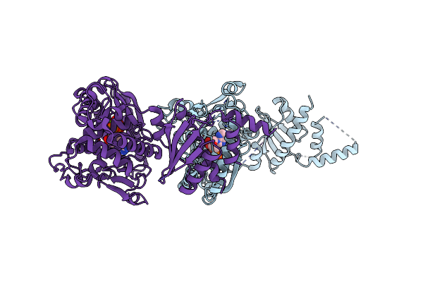
Organism: Homo sapiens
Method: X-RAY DIFFRACTION Resolution:2.70 Å Release Date: 2017-05-31 Classification: TRANSFERASE Ligands: UTP, MG, CL, EDO |
 |
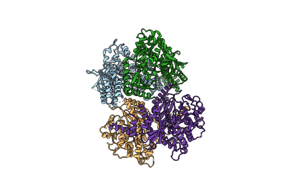
Organism: Homo sapiens
Method: X-RAY DIFFRACTION Resolution:2.80 Å Release Date: 2017-05-31 Classification: TRANSFERASE Ligands: ATP, MG, CL |
 |
Organism: Homo sapiens
Method: X-RAY DIFFRACTION Resolution:3.40 Å Release Date: 2017-05-31 Classification: TRANSFERASE |

