Search Count: 39
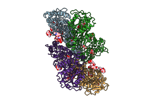 |
Organism: Rattus norvegicus
Method: ELECTRON MICROSCOPY Release Date: 2024-03-13 Classification: MEMBRANE PROTEIN Ligands: NAG, DOQ |
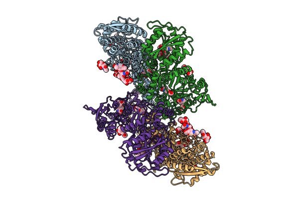 |
Organism: Rattus norvegicus
Method: ELECTRON MICROSCOPY Release Date: 2024-03-13 Classification: MEMBRANE PROTEIN Ligands: NAG, DOQ |
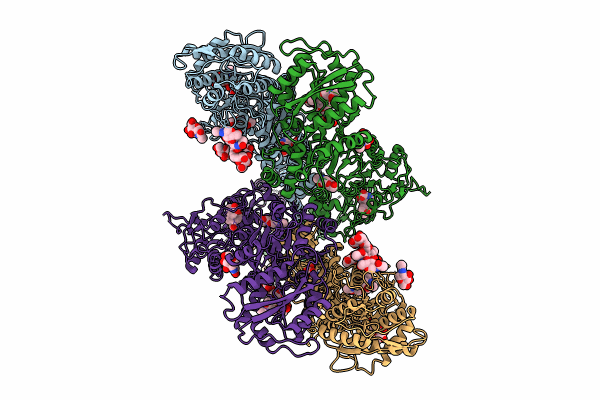 |
Organism: Rattus norvegicus
Method: ELECTRON MICROSCOPY Release Date: 2024-03-13 Classification: MEMBRANE PROTEIN Ligands: NAG, DOQ |
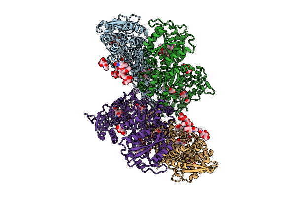 |
Organism: Rattus norvegicus
Method: ELECTRON MICROSCOPY Release Date: 2024-03-13 Classification: MEMBRANE PROTEIN Ligands: NAG, DOQ, MAN |
 |
Cryo-Em Structure Of Homomeric Kainate Receptor Gluk2 In Resting (Apo) State
Organism: Rattus norvegicus
Method: ELECTRON MICROSCOPY Release Date: 2023-11-08 Classification: MEMBRANE PROTEIN Ligands: NAG |
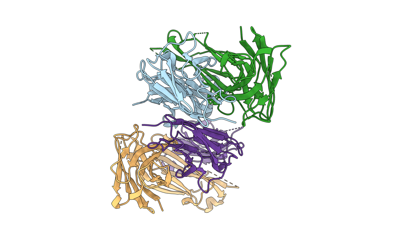 |
Organism: Mus musculus
Method: X-RAY DIFFRACTION Resolution:2.46 Å Release Date: 2022-03-02 Classification: IMMUNE SYSTEM |
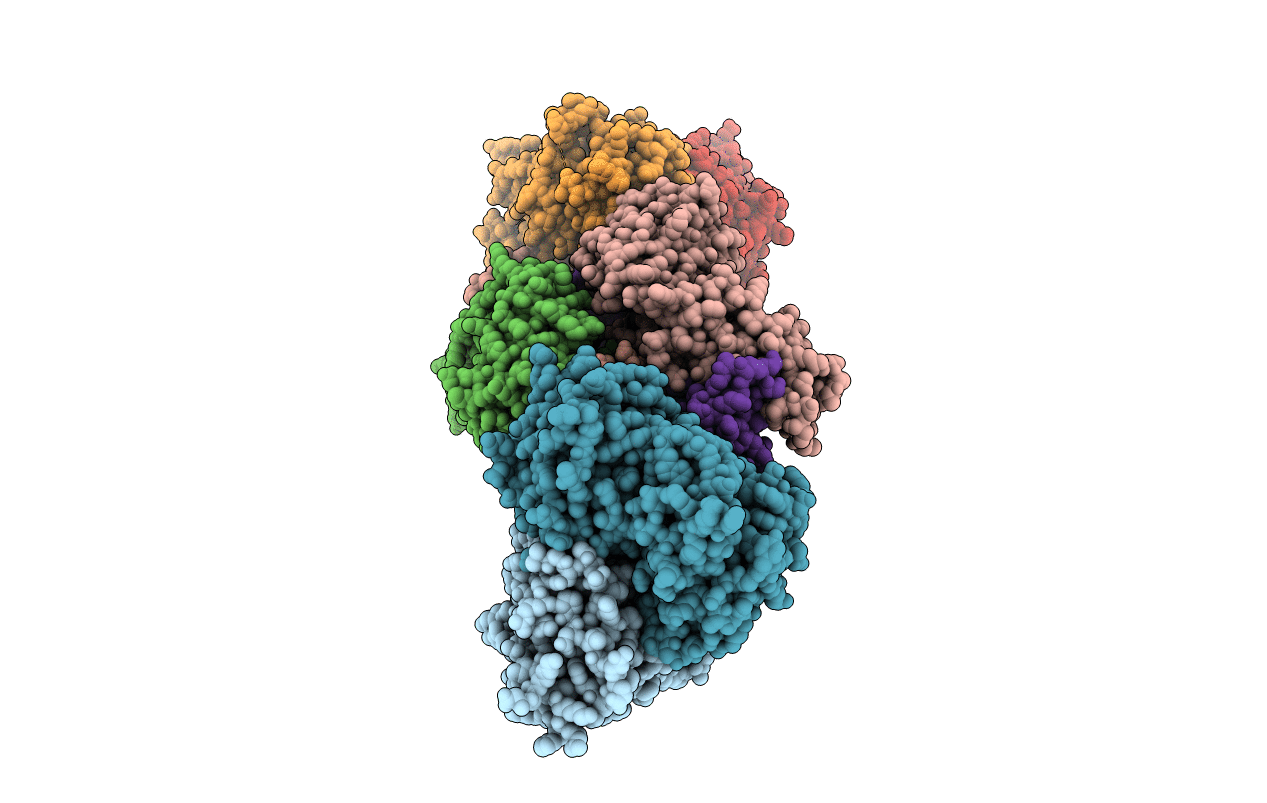 |
Organism: Xenopus laevis, Rattus norvegicus, Mus musculus
Method: X-RAY DIFFRACTION Resolution:4.55 Å Release Date: 2022-03-02 Classification: SIGNALING PROTEIN/IMMUNE SYSTEM |
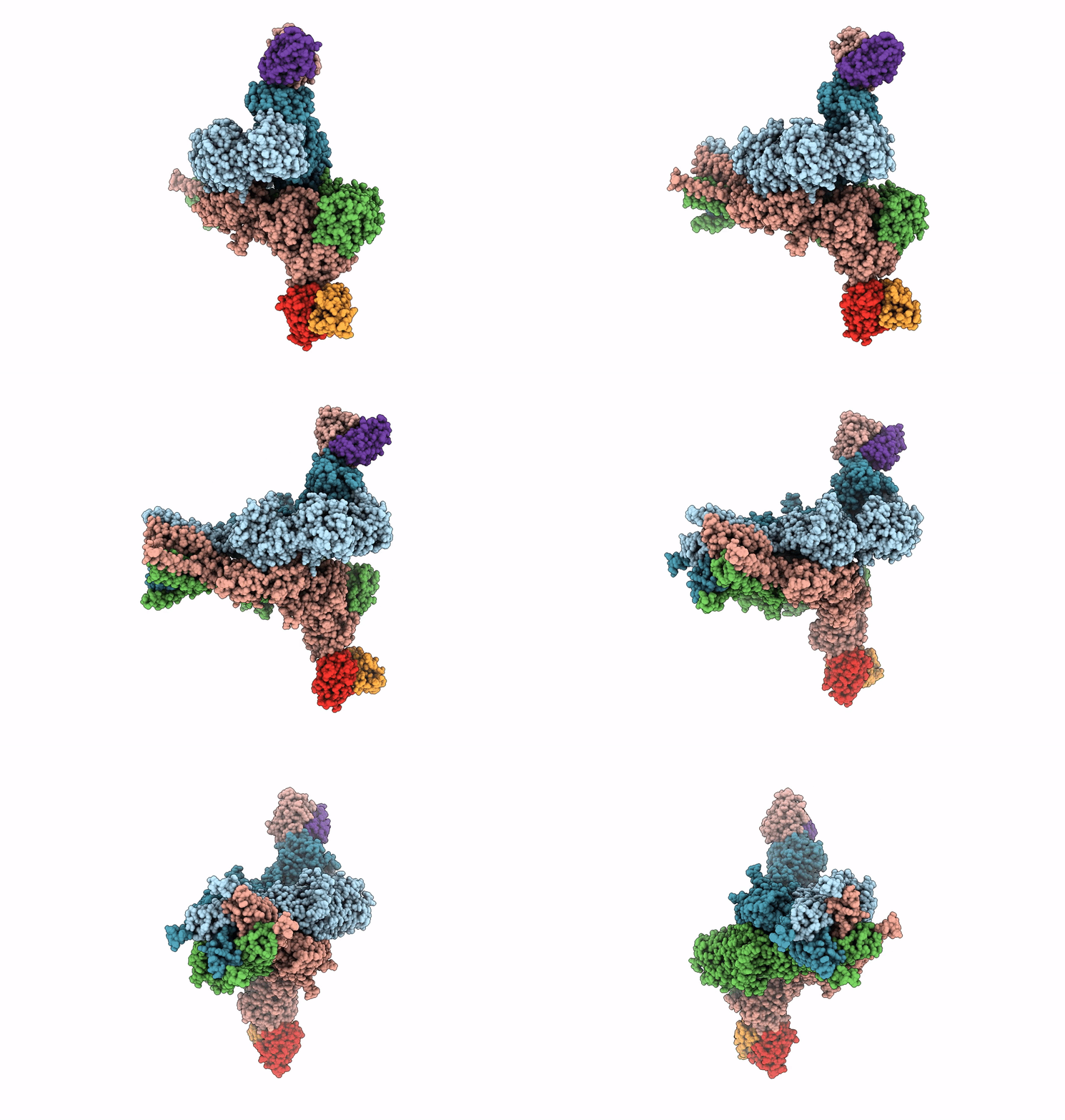 |
Organism: Rattus norvegicus, Mus musculus
Method: ELECTRON MICROSCOPY Release Date: 2022-03-02 Classification: SIGNALING PROTEIN/IMMUNE SYSTEM |
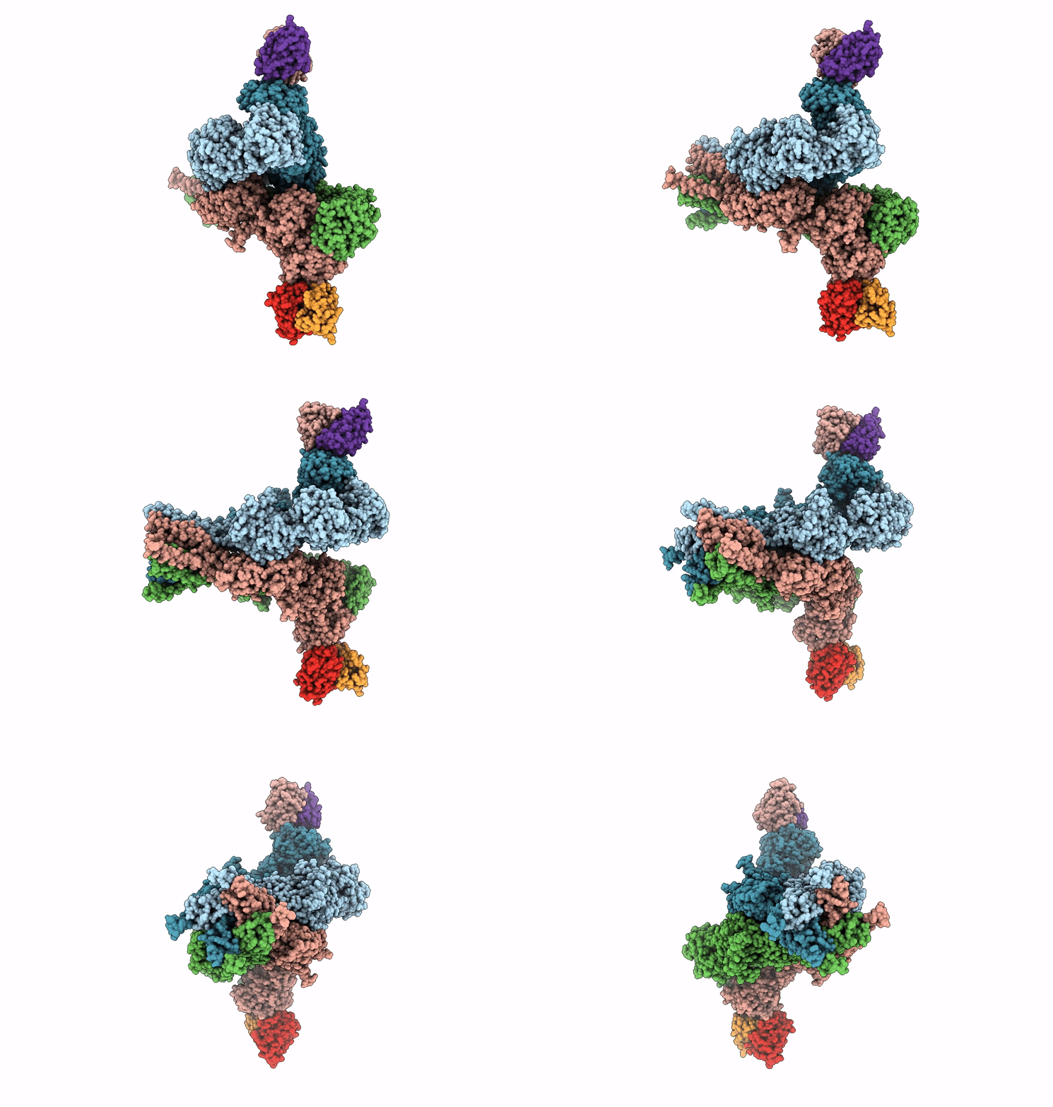 |
Organism: Rattus norvegicus, Mus musculus
Method: ELECTRON MICROSCOPY Release Date: 2022-03-02 Classification: SIGNALING PROTEIN/IMMUNE SYSTEM |
 |
Organism: Rattus norvegicus, Mus musculus
Method: ELECTRON MICROSCOPY Release Date: 2022-03-02 Classification: SIGNALING PROTEIN/IMMUNE SYSTEM |
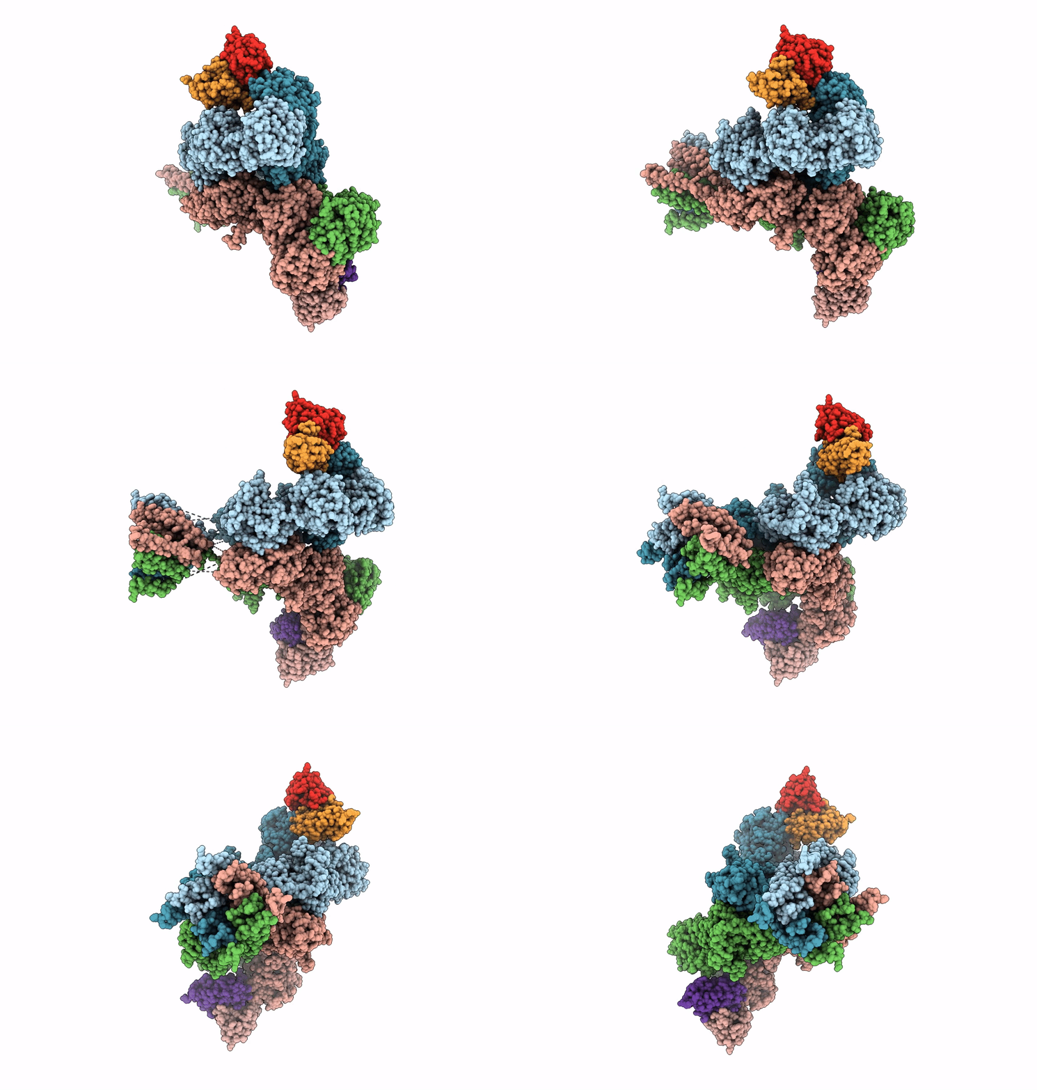 |
Cryo-Em Structure Of Glun1B-2B Nmdar In Complex With Fab5 Active Conformation
Organism: Rattus norvegicus, Mus musculus
Method: ELECTRON MICROSCOPY Release Date: 2022-03-02 Classification: SIGNALING PROTEIN/IMMUNE SYSTEM |
 |
Cryo-Em Structure Of Glun1B-2B Nmdar In Complex With Fab5 Non-Active2 Conformation
Organism: Rattus norvegicus, Mus musculus
Method: ELECTRON MICROSCOPY Release Date: 2022-03-02 Classification: SIGNALING PROTEIN/IMMUNE SYSTEM |
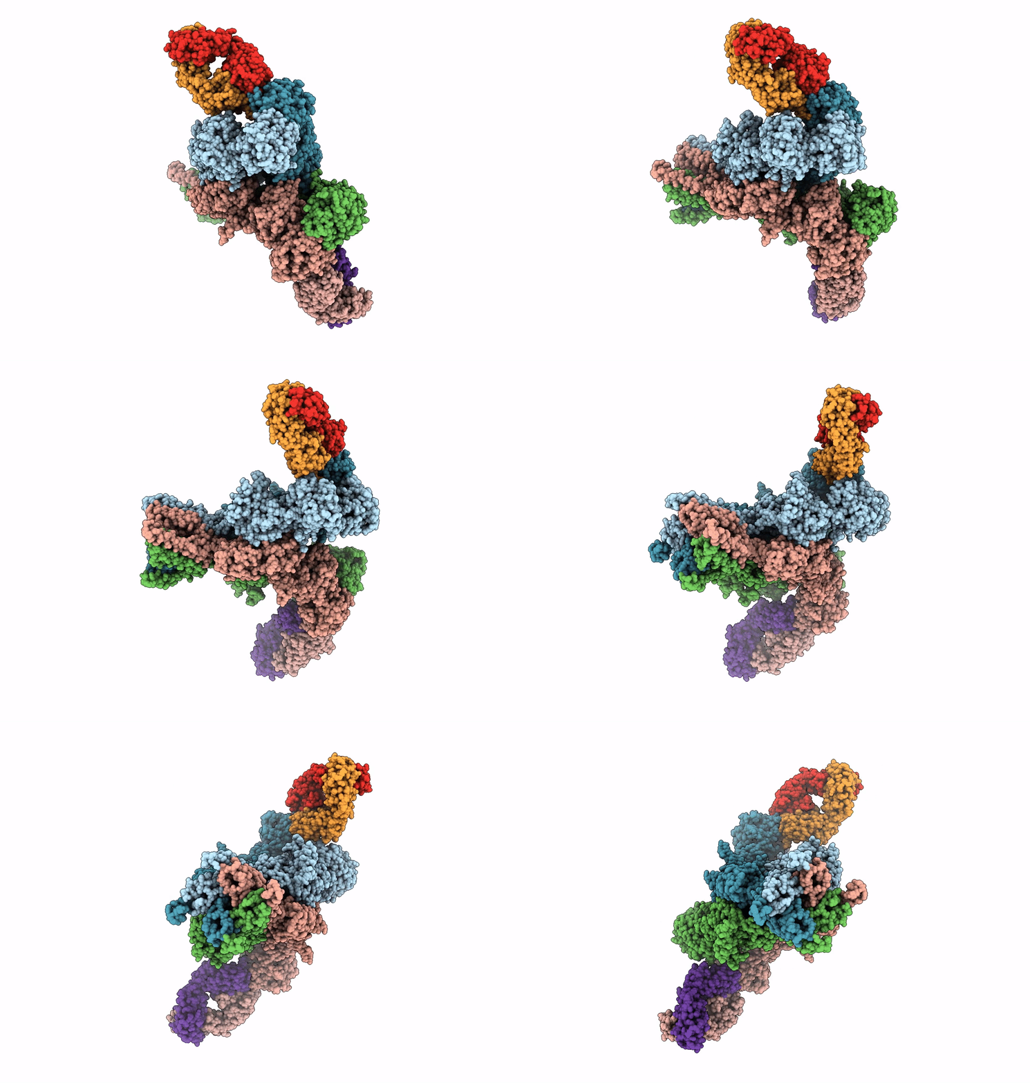 |
Cryo-Em Structure Of Glun1B-2B Nmdar In Complex With Fab5 In Non-Active1 Conformation
Organism: Rattus norvegicus, Mus musculus
Method: ELECTRON MICROSCOPY Release Date: 2022-03-02 Classification: SIGNALING PROTEIN/IMMUNE SYSTEM |
 |
Cryo-Em Structure Of Glun1B-2B Nmdar In Complex With Fab5 In Non-Active2-Like Conformation
Organism: Rattus norvegicus, Mus musculus
Method: ELECTRON MICROSCOPY Release Date: 2022-03-02 Classification: SIGNALING PROTEIN/IMMUNE SYSTEM |
 |
Glun1B-Glun2B Nmda Receptor In Non-Active 2 Conformation At 4 Angstrom Resolution
Organism: Rattus norvegicus
Method: ELECTRON MICROSCOPY Release Date: 2020-08-05 Classification: MEMBRANE PROTEIN Ligands: NAG |
 |
Glun1B-Glun2B Nmda Receptor In Non-Active 1 Conformation At 3.95 Angstrom Resolution
Organism: Rattus norvegicus
Method: ELECTRON MICROSCOPY Release Date: 2020-08-05 Classification: MEMBRANE PROTEIN Ligands: NAG |
 |
Glun1B-Glun2B Nmda Receptor In Active Conformation At 4.4 Angstrom Resolution
Organism: Rattus norvegicus
Method: ELECTRON MICROSCOPY Release Date: 2020-08-05 Classification: MEMBRANE PROTEIN Ligands: NAG |
 |
Glun1B-Glun2B Nmda Receptor In Complex With Sdz 220-040 And L689,560, Class 1
Organism: Rattus norvegicus
Method: ELECTRON MICROSCOPY Release Date: 2020-08-05 Classification: MEMBRANE PROTEIN Ligands: QGM, NAG, QGP |
 |
Glun1B-Glun2B Nmda Receptor In Complex With Sdz 220-040 And L689,560, Class 2
Organism: Rattus norvegicus
Method: ELECTRON MICROSCOPY Release Date: 2020-08-05 Classification: MEMBRANE PROTEIN Ligands: NAG, QGP, QGM |
 |
Crystal Structure Of Glun1/Glun2A Ligand-Binding Domain In Complex With L689,560 And Glutamate
Organism: Rattus norvegicus
Method: X-RAY DIFFRACTION Resolution:2.09 Å Release Date: 2020-07-15 Classification: METAL TRANSPORT Ligands: QGM, GLU |

