
Search Count: 28
 |
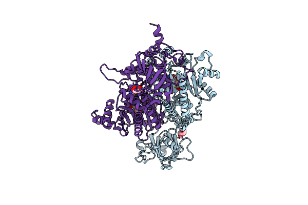
Organism: Plasmodium vivax
Method: X-RAY DIFFRACTION Release Date: 2025-06-04 Classification: LIGASE Ligands: SO4, EDO |
 |
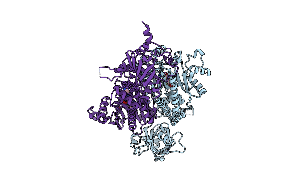
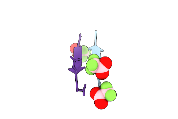
Plasmodium Vivax Aspartyl-Trna Synthetase In Complex With Aspartyl-Adenylate (Asp-Amp) Complex.
Organism: Plasmodium vivax
Method: X-RAY DIFFRACTION Release Date: 2025-06-04 Classification: LIGASE Ligands: AMO, GOL, ACT |
 |
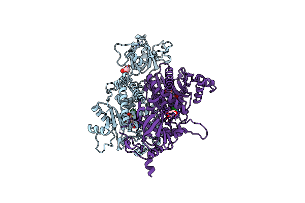
Plasmodium Vivax Aspartyl-Trna Synthetase In Complex With Aspartyl Sulfamoyl Adenosine (Asp-Ams) Complex
Organism: Plasmodium vivax
Method: X-RAY DIFFRACTION Release Date: 2025-06-04 Classification: LIGASE Ligands: DSZ, MG |
 |
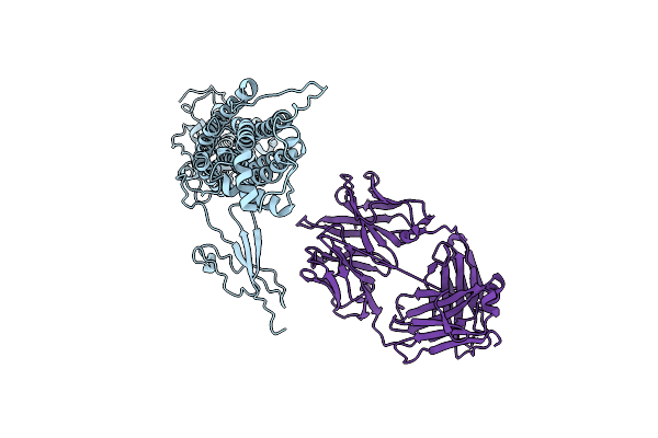
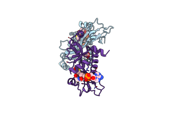
Structural Studies Of Reaction Hijacking Inhibition Of A Malaria Parasite Aspartyl-Trna Synthetase.
Organism: Plasmodium vivax
Method: X-RAY DIFFRACTION Release Date: 2025-06-04 Classification: LIGASE Ligands: A1B0L, GOL |
 |
Organism: Plasmodium falciparum 3d7
Method: X-RAY DIFFRACTION Release Date: 2024-11-27 Classification: LIGASE/INHIBITOR Ligands: A1AZG, MLI, CL, NA |
 |
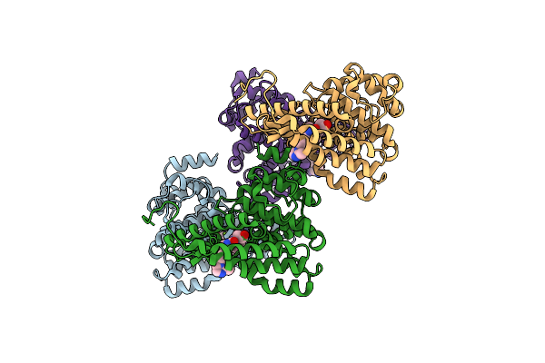
Organism: Homo sapiens, Mus musculus
Method: ELECTRON MICROSCOPY Release Date: 2021-12-22 Classification: IMMUNE SYSTEM |
 |
Aalall Segment From The Nucleoprotein Of Sars-Cov-2, Residues 217-222, Crystal Form 1
Organism: Severe acute respiratory syndrome coronavirus 2
Method: X-RAY DIFFRACTION Resolution:1.12 Å Release Date: 2021-03-17 Classification: PROTEIN FIBRIL Ligands: TFA |
 |
Aalall Segment From The Nucleoprotein Of Sars-Cov-2, Residues 217-222, Crystal Form 2
Organism: Severe acute respiratory syndrome coronavirus 2
Method: X-RAY DIFFRACTION Resolution:1.30 Å Release Date: 2021-03-17 Classification: PROTEIN FIBRIL Ligands: PG4 |
 |
Organism: Severe acute respiratory syndrome coronavirus 2
Method: X-RAY DIFFRACTION Resolution:1.10 Å Release Date: 2021-03-17 Classification: PROTEIN FIBRIL |
 |
Organism: Severe acute respiratory syndrome coronavirus 2
Method: X-RAY DIFFRACTION Resolution:1.30 Å Release Date: 2021-03-17 Classification: PROTEIN FIBRIL |
 |
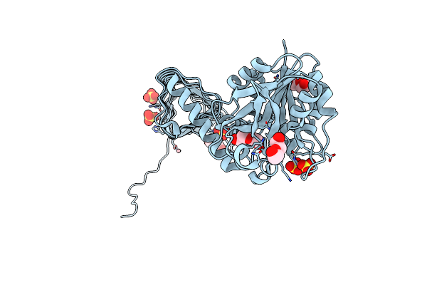
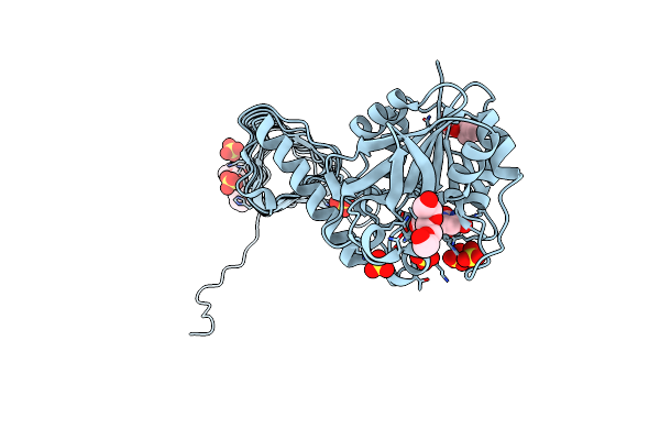
Organism: Klebsiella pneumoniae
Method: X-RAY DIFFRACTION Resolution:2.00 Å Release Date: 2017-07-19 Classification: TOXIN Ligands: ACO, ACT |
 |
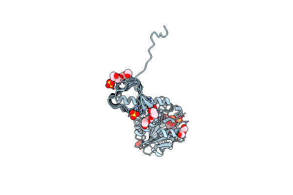
Organism: Homo sapiens
Method: X-RAY DIFFRACTION Resolution:1.72 Å Release Date: 2013-05-01 Classification: HYDROLASE/HYDROLASE INHIBITOR Ligands: ZN, MG, 1L6 |
 |
Crystal Structure Of Biotin Carboxylase From E.Coli In Complex With Potent Inhibitor 1
Organism: Escherichia coli
Method: X-RAY DIFFRACTION Resolution:2.10 Å Release Date: 2009-01-13 Classification: LIGASE Ligands: LZJ, CL |
 |
Crystal Structure Of Biotin Carboxylase From E.Coli In Complex With Potent Inhibitor 2
Organism: Escherichia coli
Method: X-RAY DIFFRACTION Resolution:2.40 Å Release Date: 2009-01-13 Classification: LIGASE Ligands: LZK |
 |
Crystal Structure Of Biotin Carboxylase From E.Coli In Complex With Potent Inhibitor 3
Organism: Escherichia coli
Method: X-RAY DIFFRACTION Resolution:2.31 Å Release Date: 2009-01-13 Classification: LIGASE Ligands: LZL, CL |
 |
Characterization Of Substrate Binding And Catalysis Of The Potential Antibacterial Target N-Acetylglucosamine-1-Phosphate Uridyltransferase (Glmu)
Organism: Haemophilus influenzae
Method: X-RAY DIFFRACTION Resolution:1.79 Å Release Date: 2008-01-15 Classification: TRANSFERASE Ligands: PEG, SO4, CO |
 |
Characterization Of Substrate Binding And Catalysis Of The Potential Antibacterial Target N-Acetylglucosamine-1-Phosphate Uridyltransferase (Glmu)
Organism: Haemophilus influenzae
Method: X-RAY DIFFRACTION Resolution:1.89 Å Release Date: 2008-01-15 Classification: TRANSFERASE Ligands: UD1, PG4, PEG, SO4 |
 |
Characterization Of Substrate Binding And Catalysis Of The Potential Antibacterial Target N-Acetylglucosamine-1-Phosphate Uridyltransferase (Glmu)
Organism: Haemophilus influenzae
Method: X-RAY DIFFRACTION Resolution:2.00 Å Release Date: 2008-01-15 Classification: TRANSFERASE Ligands: H2U, MG, PG4, PGE, SO4 |
 |
Characterization Of Substrate Binding And Catalysis Of The Potential Antibacterial Target N-Acetylglucosamine-1-Phosphate Uridyltransferase (Glmu)
Organism: Haemophilus influenzae
Method: X-RAY DIFFRACTION Resolution:2.30 Å Release Date: 2008-01-15 Classification: TRANSFERASE Ligands: UDP, PG4, PGE, SO4 |
 |
Characterization Of Substrate Binding And Catalysis Of The Potential Antibacterial Target N-Acetylglucosamine-1-Phosphate Uridyltransferase (Glmu)
Organism: Haemophilus influenzae
Method: X-RAY DIFFRACTION Resolution:2.20 Å Release Date: 2008-01-15 Classification: TRANSFERASE Ligands: URI, PG4, PGE, SO4 |