
Search Count: 466
All
Selected
 |
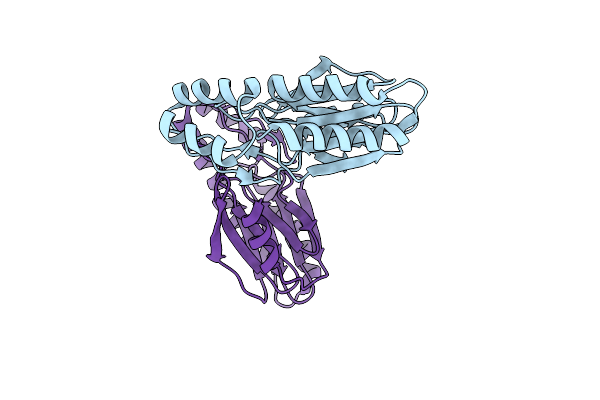
Crystal Structure Of The Double Mutant (P94A And Y128F) Of Sortase E From Thermobifida Fusca
Organism: Thermobifida fusca yx
Method: X-RAY DIFFRACTION Resolution:1.50 Å Release Date: 2026-03-11 Classification: HYDROLASE |
 |
Crystal Structure Of The Double Mutant (P94A And Y128A) Of Sortase E From Thermobifida Fusca
Organism: Thermobifida fusca yx
Method: X-RAY DIFFRACTION Resolution:1.60 Å Release Date: 2026-03-11 Classification: HYDROLASE |
 |
Crystal Structure Of The Double Mutant (P94A And C222A) Of Sortase E From Thermobifida Fusca
Organism: Thermobifida fusca yx
Method: X-RAY DIFFRACTION Resolution:1.99 Å Release Date: 2026-03-11 Classification: HYDROLASE |
 |
Crystal Structure Of The Double Mutant (Y128F And C222A) Of Sortase E From Thermobifida Fusca
Organism: Thermobifida fusca yx
Method: X-RAY DIFFRACTION Resolution:1.30 Å Release Date: 2026-03-04 Classification: HYDROLASE |
 |
Crystal Structure Of The Double Mutant (Y128A And C222A) Of Sortase E Mutant From Thermobifida Fusca
Organism: Thermobifida fusca yx
Method: X-RAY DIFFRACTION Resolution:1.50 Å Release Date: 2026-03-04 Classification: HYDROLASE |
 |
Organism: Thermobifida fusca yx
Method: X-RAY DIFFRACTION Resolution:1.69 Å Release Date: 2026-03-04 Classification: HYDROLASE |
 |
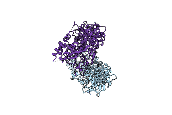
Crystal Structure Of Neisseria Gonorrhoeae Penicillin-Binding Protein 2 From Strain Fa19 Containing Six Resistance Mutations
Organism: Neisseria gonorrhoeae fa19
Method: X-RAY DIFFRACTION Resolution:2.15 Å Release Date: 2026-03-04 Classification: HYDROLASE Ligands: PO4 |
 |
Crystal Structure Of Neisseria Gonorrhoeae Penicillin-Binding Protein 2 From Strain Fa19 Containing Seven Resistance Mutations
Organism: Neisseria gonorrhoeae fa19
Method: X-RAY DIFFRACTION Resolution:1.90 Å Release Date: 2026-03-04 Classification: HYDROLASE Ligands: PO4 |
 |
Crystal Structure Of Neisseria Gonorrhoeae Penicillin-Binding Protein 2 From Strain Fa19 Containing Seven Resistance Mutations And Three Epistatic Mutations
Organism: Neisseria gonorrhoeae fa19
Method: X-RAY DIFFRACTION Resolution:1.46 Å Release Date: 2026-03-04 Classification: HYDROLASE |
 |
Organism: Staphylococcus aureus
Method: X-RAY DIFFRACTION Resolution:2.80 Å Release Date: 2026-02-18 Classification: HYDROLASE Ligands: CD, CL |
 |
Organism: Staphylococcus aureus
Method: X-RAY DIFFRACTION Resolution:3.50 Å Release Date: 2026-02-18 Classification: HYDROLASE Ligands: CD, CL |
 |
Crystal Structure Of The Transpeptidase Domain Of Pbp2 From The Neisseria Gonorrhoeae Cephalosporin-Resistant Strain H041 In Complex With Boronate Inhibitor Vnrx-14079
Organism: Neisseria gonorrhoeae
Method: X-RAY DIFFRACTION Resolution:2.10 Å Release Date: 2026-02-04 Classification: LIGASE Ligands: A1C02 |
 |
Organism: Thermobifida fusca yx
Method: X-RAY DIFFRACTION Resolution:1.50 Å Release Date: 2025-08-06 Classification: HYDROLASE |
 |
Organism: Thermobifida fusca yx
Method: X-RAY DIFFRACTION Resolution:1.83 Å Release Date: 2025-07-23 Classification: HYDROLASE Ligands: CL, NA, GOL |
 |
Organism: Thermobifida fusca yx
Method: X-RAY DIFFRACTION Resolution:1.80 Å Release Date: 2025-07-09 Classification: HYDROLASE Ligands: EDO, CL, NA |
 |
Crystal Structure Of The Transpeptidase Domain Of Pbp2 From The Neisseria Gonorrhoeae Cephalosporin Decreased Susceptibility Strain 35/02 In Complex With Boronate Inhibitor Vnrx-6884
Organism: Neisseria gonorrhoeae 35/02
Method: X-RAY DIFFRACTION Resolution:1.89 Å Release Date: 2025-01-22 Classification: LIGASE Ligands: A1BJC |
 |
Crystal Structure Of The Transpeptidase Domain Of Pbp2 From The Neisseria Gonorrhoeae Cephalosporin Decreased Susceptibility Strain 35/02 In Complex With Boronate Inhibitor Vnrx-6752
Organism: Neisseria gonorrhoeae 35/02
Method: X-RAY DIFFRACTION Resolution:2.61 Å Release Date: 2025-01-22 Classification: LIGASE Ligands: A1BJB |
 |
Connectase T1A C192S Mutant From Methanocaldococcus Mazei With Peptide Substrate
Organism: Methanosarcina mazei
Method: X-RAY DIFFRACTION Resolution:1.98 Å Release Date: 2025-01-15 Classification: LIGASE |
 |
Organism: Methanosarcina mazei
Method: X-RAY DIFFRACTION Resolution:3.40 Å Release Date: 2024-12-25 Classification: LIGASE |
 |
Organism: Acinetobacter baumannii
Method: X-RAY DIFFRACTION Resolution:3.31 Å Release Date: 2024-12-04 Classification: HYDROLASE |