
Search Count: 42,062
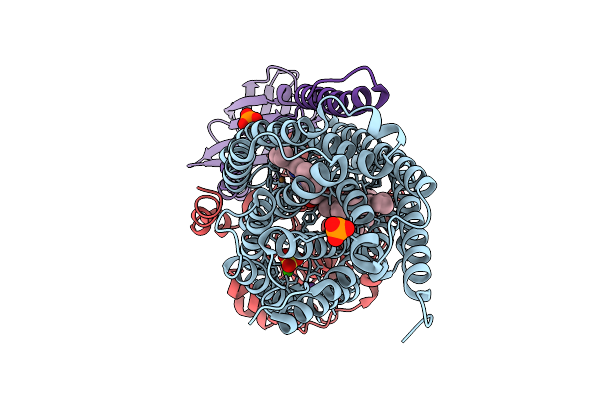 |
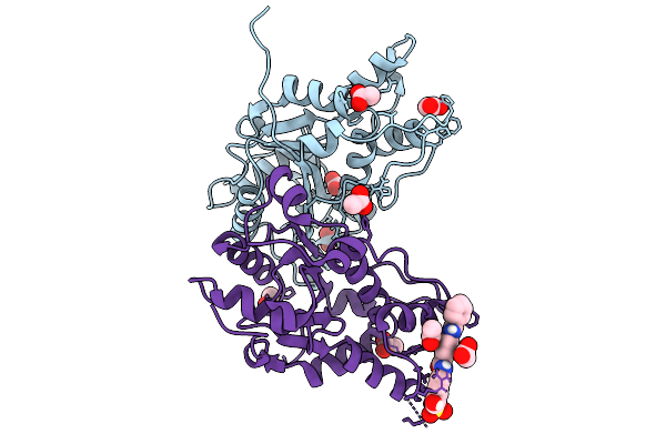
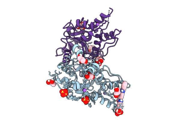
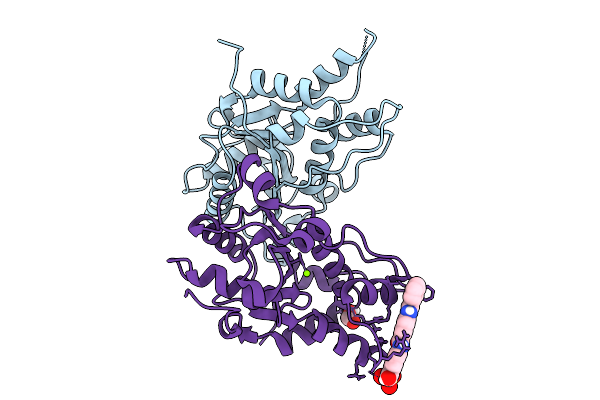
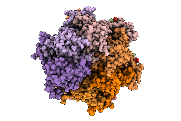
Organism: Stutzerimonas stutzeri atcc 14405 = ccug 16156
Method: X-RAY DIFFRACTION Resolution:2.90 Å Release Date: 2026-01-28 Classification: OXIDOREDUCTASE Ligands: HEM, CU, CA, PO4, NA, SO4, HEC |
 |
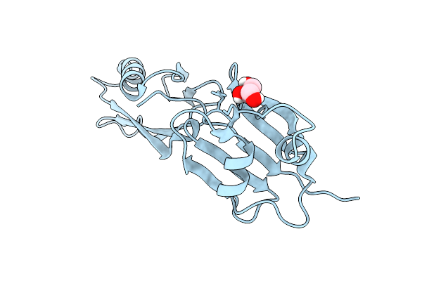
The Apo-Mtrex1 Crystal Structure For Soaking Experiments (Soaking Condition 1)
Organism: Mus musculus
Method: X-RAY DIFFRACTION Resolution:1.70 Å Release Date: 2026-01-28 Classification: HYDROLASE Ligands: ACT |
 |
The Apo-Mtrex1 Crystal Structure For Soaking Experiments (Soaking Condition 2)
Organism: Mus musculus
Method: X-RAY DIFFRACTION Resolution:1.40 Å Release Date: 2026-01-28 Classification: HYDROLASE Ligands: ACT |
 |
The Apo-Mtrex1 Crystal Structure For Soaking Experiments (Soaking Condition 3)
Organism: Mus musculus
Method: X-RAY DIFFRACTION Resolution:1.60 Å Release Date: 2026-01-28 Classification: HYDROLASE Ligands: GOL |
 |
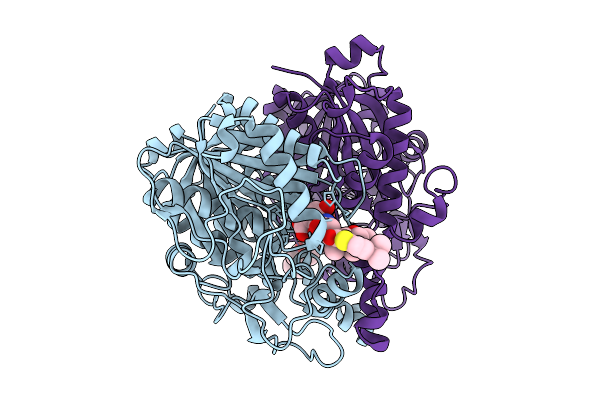
The Mtrex1-Nsc 37203 Complex Structure By Co-Crystallization (Nsc 37203 Complex 1)
Organism: Mus musculus
Method: X-RAY DIFFRACTION Resolution:1.91 Å Release Date: 2026-01-28 Classification: HYDROLASE Ligands: DMS, LI, EPE, SO4, A1L5X |
 |
The Mtrex1-Nsc 37203 Complex Structure By Soaking In Soaking Condition 1 (Nsc 37203 Complex 2)
Organism: Mus musculus
Method: X-RAY DIFFRACTION Resolution:1.80 Å Release Date: 2026-01-28 Classification: HYDROLASE Ligands: DMS, MG, ACT, A1L5X, CAC, NA |
 |
The Mtrex1-Nsc 37203 Complex Structure By Co-Crystallization (Nsc 37203 Complex 3)
Organism: Mus musculus
Method: X-RAY DIFFRACTION Resolution:1.70 Å Release Date: 2026-01-28 Classification: HYDROLASE Ligands: SO4, A1L5X |
 |
The Mtrex1-Nsc 37204 Complex Structure By Soaking In Soaking Condition 2 (Nsc 37204 Complex 1)
Organism: Mus musculus
Method: X-RAY DIFFRACTION Resolution:1.90 Å Release Date: 2026-01-28 Classification: HYDROLASE Ligands: ACT, A1L5Z |
 |
The Mtrex1-Nsc 37204 Complex Structure By Soaking In Soaking Condition 3 (Nsc 37204 Complex 2)
Organism: Mus musculus
Method: X-RAY DIFFRACTION Resolution:1.70 Å Release Date: 2026-01-28 Classification: HYDROLASE Ligands: A1L5Z, GOL, SO4, NH4 |
 |
The Mtrex1-Nsc 37204 Complex Structure By Soaking In Soaking Condition 1 (Nsc 37204 Complex 3)
Organism: Mus musculus
Method: X-RAY DIFFRACTION Resolution:1.70 Å Release Date: 2026-01-28 Classification: HYDROLASE Ligands: ACT, A1L5Z |
 |
The Mtrex1-Nsc 37215 Complex Structure By Co-Crystallization (Nsc 37215-1 Complex 1)
Organism: Mus musculus
Method: X-RAY DIFFRACTION Resolution:1.70 Å Release Date: 2026-01-28 Classification: HYDROLASE Ligands: SIN, DMS, A1L50, MG |
 |
The Mtrex1-Nsc 37215 Complex Structure By Soaking In Soaking Condition 2 (Nsc 37215-1 Complex 2)
Organism: Mus musculus
Method: X-RAY DIFFRACTION Resolution:2.00 Å Release Date: 2026-01-28 Classification: HYDROLASE Ligands: ACT, A1L50 |
 |
The Mtrex1-Nsc 37215 Complex Structure By Soaking In Soaking Condition 1 (Nsc 37215-2 Complex 1)
Organism: Mus musculus
Method: X-RAY DIFFRACTION Resolution:1.90 Å Release Date: 2026-01-28 Classification: HYDROLASE Ligands: ACT, A1L50 |
 |
The Mtrex1-Nsc 37215 Complex Structure By Co-Crystallization (Nsc 37215-2 Complex 2)
Organism: Mus musculus
Method: X-RAY DIFFRACTION Resolution:1.80 Å Release Date: 2026-01-28 Classification: HYDROLASE |
 |
The Mtrex1-Nsc 37215 Complex Structure By Co-Crystallization (Nsc 37215-2 Complex 3)
Organism: Mus musculus
Method: X-RAY DIFFRACTION Resolution:1.70 Å Release Date: 2026-01-28 Classification: HYDROLASE Ligands: A1L50, MG, FMT |
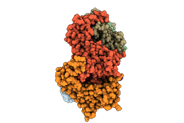 |
Pentamer Msp1 From S.Cerevisiae (With A Catalytic Dead Mutation) In Complex With An Unknown Peptide Substrate State2
Organism: Saccharomyces cerevisiae s288c
Method: ELECTRON MICROSCOPY Release Date: 2026-01-28 Classification: MEMBRANE PROTEIN Ligands: ATP, MG |
 |
Sixteen Polymer Msp1 From S.Cerevisiae (With A Catalytic Dead Mutation) In Complex With An Unknown Peptide Substrate
Organism: Saccharomyces cerevisiae s288c, Escherichia coli bl21(de3)
Method: ELECTRON MICROSCOPY Release Date: 2026-01-28 Classification: MEMBRANE PROTEIN Ligands: ATP, MG |
 |
Twenty-Two Polymer Msp1 From S.Cerevisiae(With A Catalytic Dead Mutation) In Complex With An Unknown Peptide Substrate
Organism: Saccharomyces cerevisiae s288c, Escherichia coli bl21(de3)
Method: ELECTRON MICROSCOPY Release Date: 2026-01-28 Classification: MEMBRANE PROTEIN Ligands: MG, ATP |
 |
Organism: Escherichia coli k-12
Method: X-RAY DIFFRACTION Resolution:2.70 Å Release Date: 2026-01-28 Classification: TRANSFERASE Ligands: A1EWE |
 |
Organism: Myxococcus xanthus dz2
Method: X-RAY DIFFRACTION Resolution:1.90 Å Release Date: 2026-01-28 Classification: TRANSLOCASE Ligands: GOL |