
Search Count: 134
All
Selected
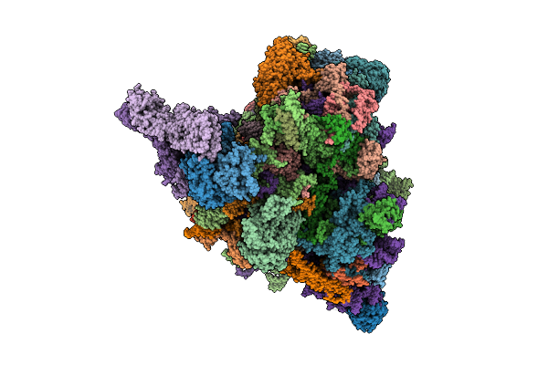 |
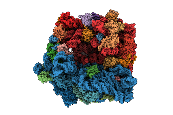
Translational Activators Aep1, Aep2 And Atp25 In Complex With Mrna And The Yeast Mitochondrial Ribosome
Organism: Saccharomyces cerevisiae w303
Method: ELECTRON MICROSCOPY Release Date: 2025-12-17 Classification: RIBOSOME Ligands: MG, GTP |
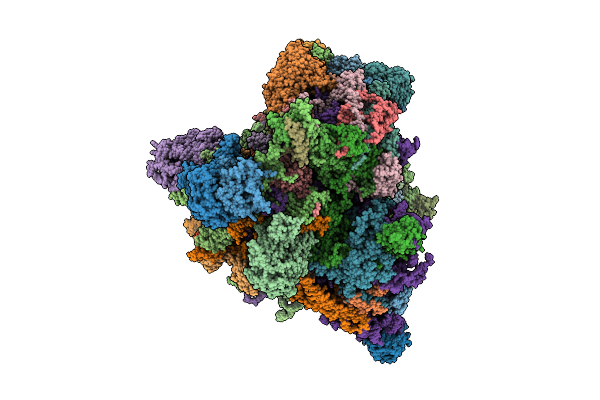 |
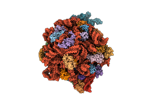
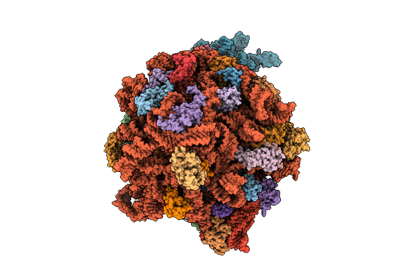
Translational Activator Aep3 In Complex With Mrna And The Yeast Mitochondrial Ribosome
Organism: Saccharomyces cerevisiae w303, Saccharomyces cerevisiae
Method: ELECTRON MICROSCOPY Release Date: 2025-12-17 Classification: RIBOSOME Ligands: MG, GTP |
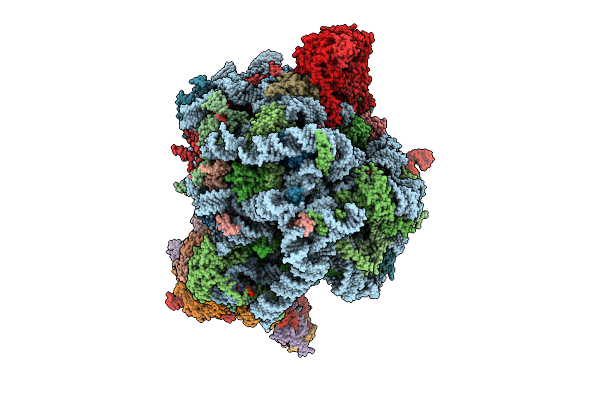 |
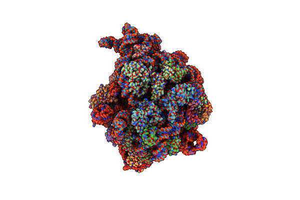
Human Quaternary Complex Of A Translating 80S Ribosome, Nac, Metap1 And Natd
Organism: Homo sapiens, Saccharomyces cerevisiae s288c, Aequorea victoria, Brachypodium distachyon
Method: ELECTRON MICROSCOPY Release Date: 2025-12-17 Classification: RIBOSOME Ligands: ZN, COA, GTP |
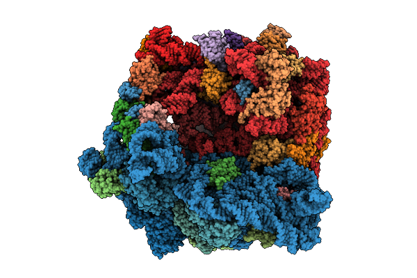 |
Structure Of The Bacterial Ribosome Without Hypoxia-Induced Rrna Modifications
Organism: Escherichia phage t4, Escherichia coli bw25113
Method: ELECTRON MICROSCOPY Release Date: 2025-11-12 Classification: RIBOSOME Ligands: MG |
 |
Structure Of The Bacterial Ribosome With Hypoxia-Induced Rrna Modifications
Organism: Escherichia phage t4, Escherichia coli bw25113
Method: ELECTRON MICROSCOPY Release Date: 2025-11-12 Classification: RIBOSOME Ligands: MG |
 |
Organism: Escherichia coli
Method: ELECTRON MICROSCOPY Release Date: 2025-10-22 Classification: RIBOSOME Ligands: ZN, PRO |
 |
Structure Of E.Coli Ribosome In Complex With An Engineered Arrest Peptide And Trigger Factor
Organism: Escherichia coli
Method: ELECTRON MICROSCOPY Release Date: 2025-10-22 Classification: RIBOSOME Ligands: ZN, PRO |
 |
Organism: Oryctolagus cuniculus, Homo sapiens
Method: ELECTRON MICROSCOPY Release Date: 2025-07-23 Classification: TRANSLATION |
 |
Organism: Escherichia coli
Method: ELECTRON MICROSCOPY Release Date: 2025-06-18 Classification: RIBOSOME Ligands: ZN, PRO |
 |
Structure Of E.Coli Ribosome In Complex With An Engineered Arrest Peptide And Trigger Factor
Organism: Escherichia coli
Method: ELECTRON MICROSCOPY Release Date: 2025-06-18 Classification: RIBOSOME Ligands: ZN, PRO |
 |
Method: ELECTRON MICROSCOPY
Release Date: 2025-05-28 Classification: RIBOSOME Ligands: MG, SPD, PUT, SPM, ZN |
 |
Method: ELECTRON MICROSCOPY
Release Date: 2025-05-28 Classification: RIBOSOME Ligands: MG, SPD, SPM, PUT, ZN |
 |
Structure Of Mycobacterium Smegmatis 50S Ribosomal Subunit Bound To Hflx And Erythromycin:50S-Hflx-B-Ery
Organism: Mycolicibacterium smegmatis mc2 155
Method: ELECTRON MICROSCOPY Release Date: 2025-04-30 Classification: RIBOSOME Ligands: GCP, ERY |
 |
E. Coli Pre-Elongation Complex Without An A-Site Trna With Eq2-Ybit In Non-Hydrolytic 1/Ptim(A) Conformation
Organism: Escherichia coli
Method: ELECTRON MICROSCOPY Release Date: 2025-04-02 Classification: RIBOSOME Ligands: MG, ZN, ATP, NA |
 |
E. Coli Pre-Elongation Complex Without An A-Site Trna With Eq2-Etta In Hydrolytic 1 Conformation
Organism: Escherichia coli
Method: ELECTRON MICROSCOPY Release Date: 2025-04-02 Classification: RIBOSOME Ligands: MG, ZN, ATP, NA |
 |
Organism: Escherichia coli
Method: ELECTRON MICROSCOPY Release Date: 2025-04-02 Classification: RIBOSOME Ligands: MG, ATP, NA, FME |
 |
Organism: Escherichia coli
Method: ELECTRON MICROSCOPY Release Date: 2025-04-02 Classification: RIBOSOME Ligands: MG, ATP, NA, FME |
 |
Organism: Mycolicibacterium smegmatis mc2 155
Method: ELECTRON MICROSCOPY Release Date: 2025-02-19 Classification: RIBOSOME |
 |
Structure Of Mycobacterium Smegmatis 50S Ribosomal Subunit Bound To Hflx:50S-Hflx-A
Organism: Mycolicibacterium smegmatis mc2 155
Method: ELECTRON MICROSCOPY Release Date: 2025-02-19 Classification: RIBOSOME Ligands: GCP |
 |
Structure Of Mycobacterium Smegmatis 50S Ribosomal Subunit Bound To Hflx:50S-Hflx-B
Organism: Mycolicibacterium smegmatis mc2 155
Method: ELECTRON MICROSCOPY Release Date: 2025-02-19 Classification: RIBOSOME Ligands: GCP |