
Search Count: 83,600
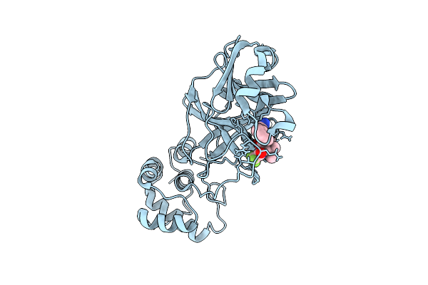 |
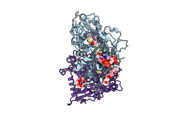
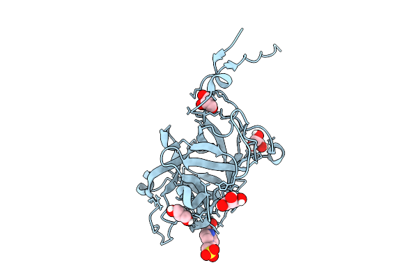
X-Ray Structure Of Sars-Cov-2 Main Protease V186G Covalently Bound To Compound Grl-051-22 At 1.3 A
Organism: Severe acute respiratory syndrome coronavirus 2
Method: X-RAY DIFFRACTION Release Date: 2025-11-26 Classification: VIRAL PROTEIN, HYDROLASE Ligands: A1BFE |
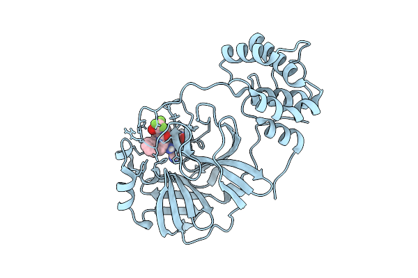 |
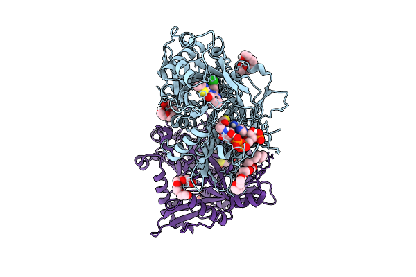
X-Ray Structure Of Sars-Cov-2 Main Protease Covalently Bound To Compound Grl-051-22 At 1.75 A.
Organism: Severe acute respiratory syndrome coronavirus 2
Method: X-RAY DIFFRACTION Release Date: 2025-11-26 Classification: VIRAL PROTEIN, HYDROLASE Ligands: A1BFE |
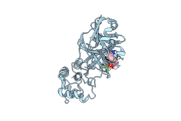 |
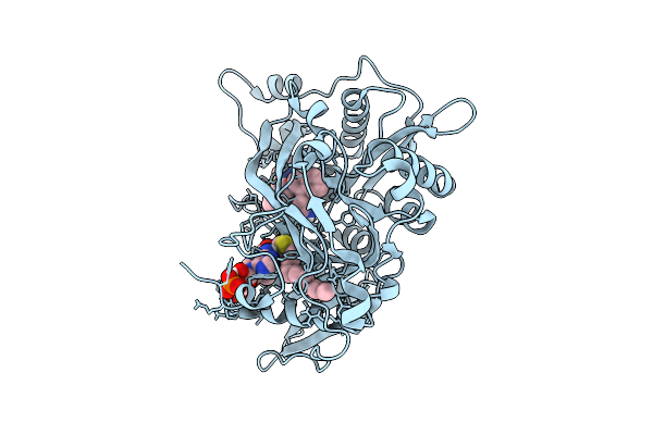
X-Ray Structure Of Sars-Cov-2 Main Protease T190I Covalently Bound To Compound Grl-051-22 At 1.5 A
Organism: Severe acute respiratory syndrome coronavirus
Method: X-RAY DIFFRACTION Release Date: 2025-11-26 Classification: VIRAL PROTEIN, HYDROLASE Ligands: A1BFE, NA |
 |
Organism: Xanthomonas citri
Method: X-RAY DIFFRACTION Release Date: 2025-11-26 Classification: TRANSFERASE Ligands: 12C, SO4 |
 |
Crystal Structure Of Cryptosporidium Parvum N-Myristoyltransferase With Bound Myristoyl-Coa And Inhibitor 20045
Organism: Cryptosporidium parvum iowa ii
Method: X-RAY DIFFRACTION Release Date: 2025-11-26 Classification: TRANSFERASE/INHIBITOR Ligands: MYA, A1BHP, CL, PG4, 1PE, PGE |
 |
Crystal Structure Of Cryptosporidium Parvum N-Myristoyltransferase With Bound Myristoyl-Coa And Inhibitor 20057
Organism: Cryptosporidium parvum iowa ii
Method: X-RAY DIFFRACTION Release Date: 2025-11-26 Classification: TRANSFERASE/INHIBITOR Ligands: MYA, A1BHQ, CL, 1PE, PG4 |
 |
Crystal Structure Of Cryptosporidium Parvum N-Myristoyltransferase With Bound Myristoyl-Coa And Inhibitor 20084
Organism: Cryptosporidium parvum iowa ii
Method: X-RAY DIFFRACTION Release Date: 2025-11-26 Classification: TRANSFERASE/INHIBITOR Ligands: MYA, A1BHR, CL |
 |
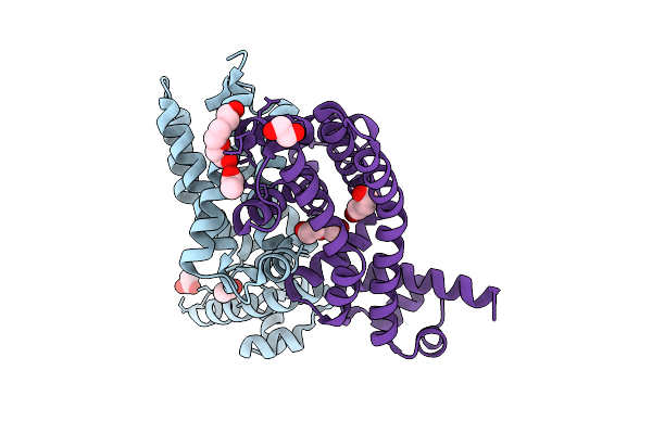
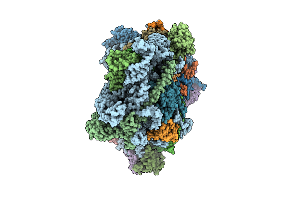
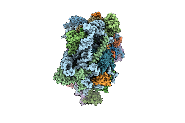
Organism: Orthopoxvirus vaccinia
Method: ELECTRON MICROSCOPY Release Date: 2025-11-26 Classification: TRANSCRIPTION Ligands: MG, ZN |
 |
Structure Of The Saccharomyces Cerevisiae Pmt4-Mir Domain With Bound Ligands
Organism: Saccharomyces cerevisiae
Method: X-RAY DIFFRACTION Release Date: 2025-11-26 Classification: PEPTIDE BINDING PROTEIN Ligands: EPE, GOL |
 |
Structure Of The Chaetomium Thermophilum Pmt4-Mir Domain With Bound Ligands
Organism: Chaetomium
Method: X-RAY DIFFRACTION Release Date: 2025-11-26 Classification: PEPTIDE BINDING PROTEIN Ligands: EDO, ACT, NA |
 |
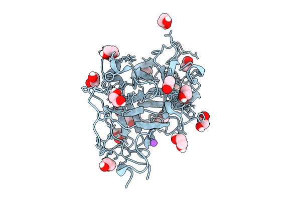
Organism: Escherichia coli k-12
Method: X-RAY DIFFRACTION Release Date: 2025-11-26 Classification: TRANSCRIPTION Ligands: ACT, PEG, CL, PGE, EDO |
 |
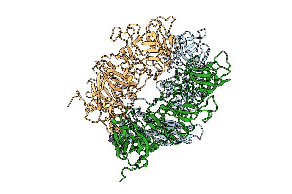
Organism: Rabbit hemorrhagic disease virus
Method: ELECTRON MICROSCOPY Release Date: 2025-11-26 Classification: VIRUS LIKE PARTICLE |
 |
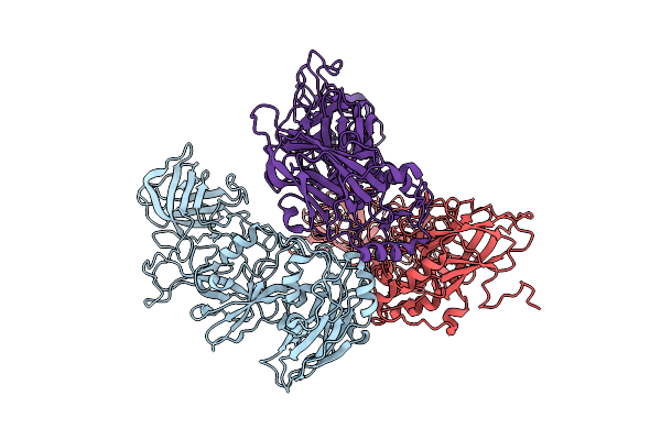
Organism: Rabbit hemorrhagic disease virus-ast89, Oryctolagus cuniculus
Method: ELECTRON MICROSCOPY Release Date: 2025-11-26 Classification: VIRUS LIKE PARTICLE |
 |
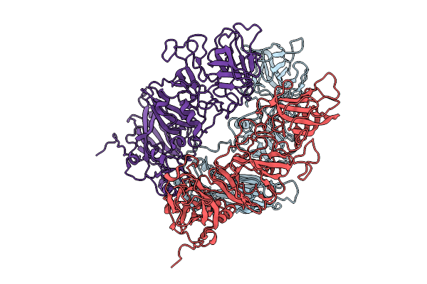
Organism: Rabbit hemorrhagic disease virus
Method: ELECTRON MICROSCOPY Release Date: 2025-11-26 Classification: VIRAL PROTEIN |
 |
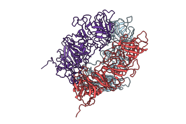
Organism: Rabbit hemorrhagic disease virus
Method: ELECTRON MICROSCOPY Release Date: 2025-11-26 Classification: VIRUS |
 |
Organism: Oryctolagus cuniculus
Method: ELECTRON MICROSCOPY Release Date: 2025-11-26 Classification: VIRUS |
 |
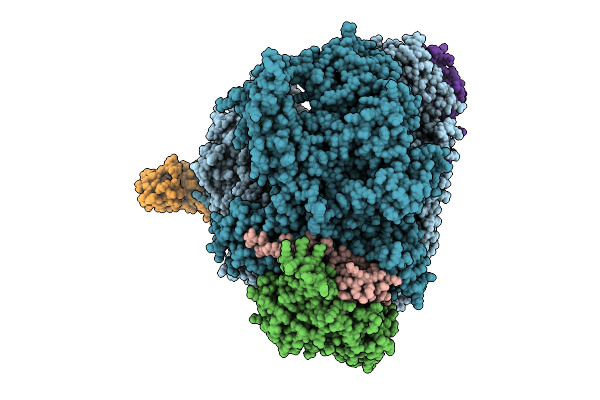
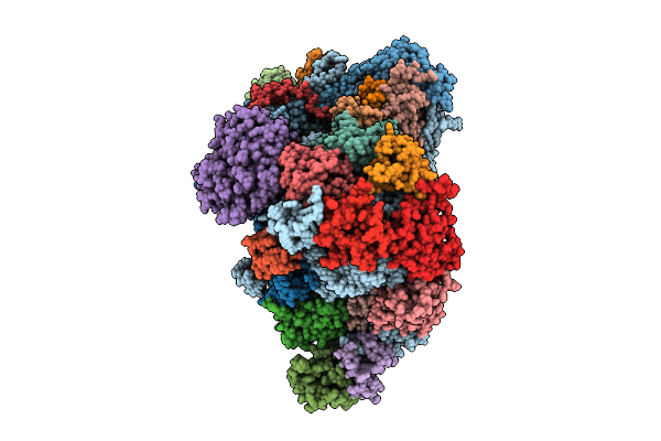
Organism: Homo sapiens, Sars bat coronavirus
Method: ELECTRON MICROSCOPY Release Date: 2025-11-26 Classification: RIBOSOME |
 |
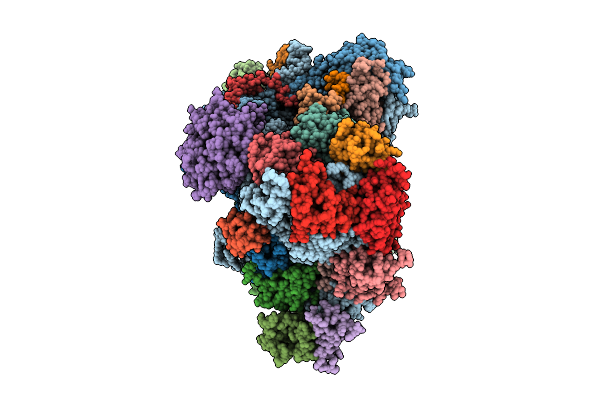
Organism: Homo sapiens, Middle east respiratory syndrome-related coronavirus
Method: ELECTRON MICROSCOPY Release Date: 2025-11-26 Classification: RIBOSOME |
 |
Bat Mersr-Cov Nl140422 Nsp1 Bound To The Human 40S Ribosomal Subunit-State1
Organism: Homo sapiens, Middle east respiratory syndrome-related coronavirus
Method: ELECTRON MICROSCOPY Release Date: 2025-11-26 Classification: RIBOSOME |
 |
Bat Mersr-Cov Nl140422 Nsp1 Bound To The Human 40S Ribosomal Subunit-State2
Organism: Homo sapiens, Middle east respiratory syndrome-related coronavirus
Method: ELECTRON MICROSCOPY Release Date: 2025-11-26 Classification: RIBOSOME |