
Search Count: 80,749
 |
Organism: Severe acute respiratory syndrome coronavirus
Method: X-RAY DIFFRACTION Release Date: 2025-12-03 Classification: VIRAL PROTEIN Ligands: A1AWG |
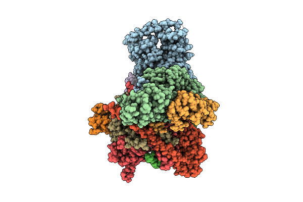 |
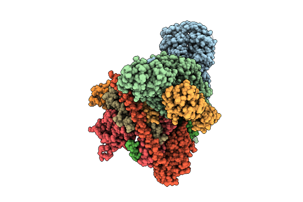
Cryo-Em Structure Of Cop9 Signalosome Precatalytic State With Neddylated Cullin-1
Organism: Homo sapiens
Method: ELECTRON MICROSCOPY Release Date: 2025-12-03 Classification: SIGNALING PROTEIN Ligands: ZN |
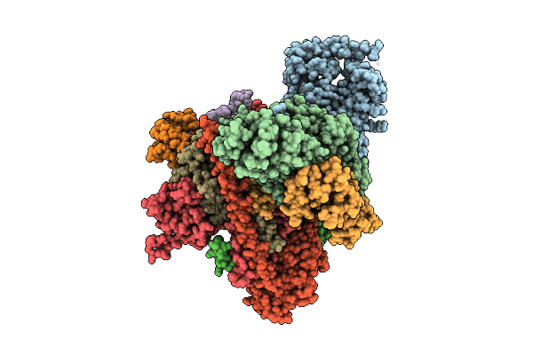 |
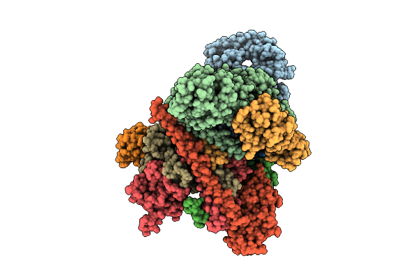
Cryo-Em Structure Of Cop9 Signalosome Precatalytic State With Neddylated Cullin-2
Organism: Homo sapiens
Method: ELECTRON MICROSCOPY Release Date: 2025-12-03 Classification: SIGNALING PROTEIN Ligands: ZN |
 |
Organism: Homo sapiens
Method: ELECTRON MICROSCOPY Release Date: 2025-12-03 Classification: SIGNALING PROTEIN Ligands: 6LT, ZN |
 |
Organism: Homo sapiens
Method: ELECTRON MICROSCOPY Release Date: 2025-12-03 Classification: SIGNALING PROTEIN Ligands: ZN |
 |
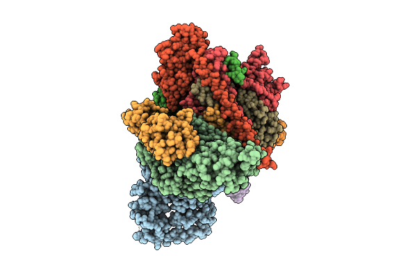
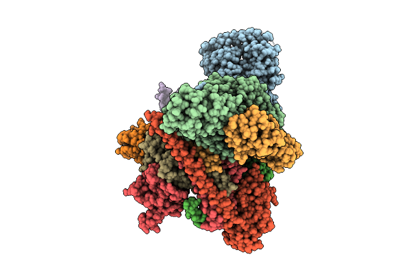
Cryo-Em Structure Of Cop9 Signalosome Precatalytic State With Neddylated Cullin-4A
Organism: Homo sapiens
Method: ELECTRON MICROSCOPY Release Date: 2025-12-03 Classification: SIGNALING PROTEIN Ligands: ZN |
 |
Cryo-Em Structure Of Cop9 Signalosome Precatalytic State With Neddylated Cullin-3
Organism: Homo sapiens
Method: ELECTRON MICROSCOPY Release Date: 2025-12-03 Classification: SIGNALING PROTEIN Ligands: ZN |
 |
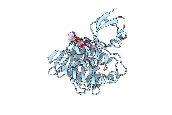
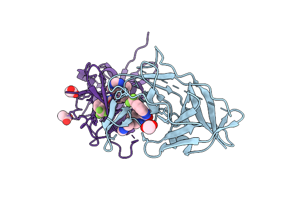
A Rare Open Conformation For Ubl2 Domain Of Papain-Like Protease Of Sars-Cov2
Organism: Severe acute respiratory syndrome coronavirus 2
Method: X-RAY DIFFRACTION Release Date: 2025-12-03 Classification: VIRAL PROTEIN Ligands: GOL, ZN, CL |
 |
A Rare Open Conformation For Ubl2 Domain Of Papain-Like Protease Without Zinc Of Sars-Cov2
Organism: Severe acute respiratory syndrome coronavirus 2
Method: X-RAY DIFFRACTION Release Date: 2025-12-03 Classification: VIRAL PROTEIN Ligands: GOL, ZN, CL, SO4 |
 |
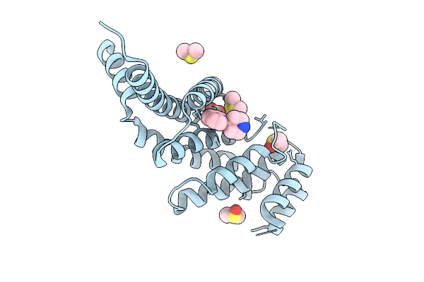
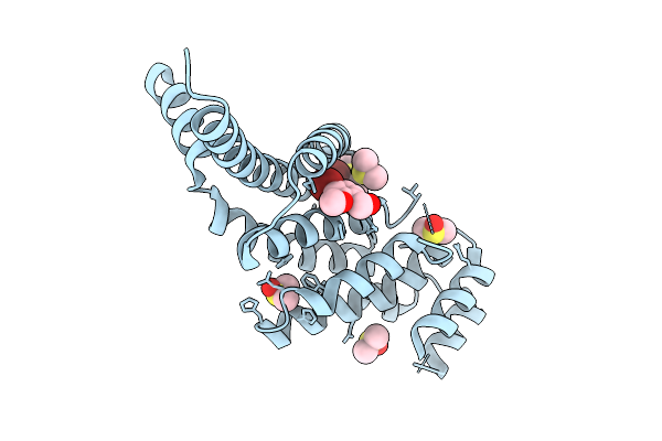
Organism: Homo sapiens
Method: X-RAY DIFFRACTION Release Date: 2025-12-03 Classification: PROTEIN BINDING Ligands: GOL, A1IU1, DMS |
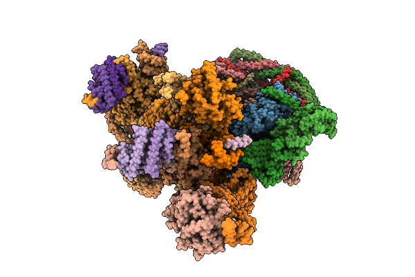 |
Organism: Homo sapiens, Sus scrofa
Method: ELECTRON MICROSCOPY Release Date: 2025-12-03 Classification: MOTOR PROTEIN Ligands: ADP, ATP, ZN |
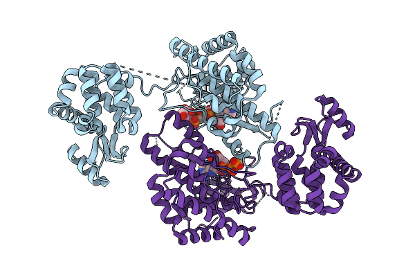 |
Crystal Structure Of Phosphatidyl Inositol 4-Kinase Ii Beta In Complex With Hh5129
Organism: Homo sapiens, Enterobacteria phage t4
Method: X-RAY DIFFRACTION Release Date: 2025-12-03 Classification: TRANSFERASE Ligands: A1IVA |
 |
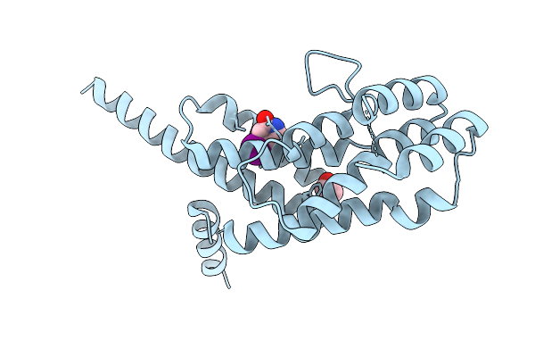
Organism: Homo sapiens
Method: X-RAY DIFFRACTION Release Date: 2025-12-03 Classification: PROTEIN BINDING Ligands: GOL, A1IV7, DMS |
 |
Organism: Homo sapiens
Method: X-RAY DIFFRACTION Release Date: 2025-12-03 Classification: PROTEIN BINDING Ligands: HHQ, EDO |
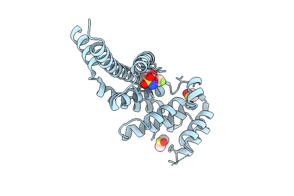 |
Organism: Homo sapiens
Method: X-RAY DIFFRACTION Release Date: 2025-12-03 Classification: PROTEIN BINDING Ligands: GOL, LL0, DMS |
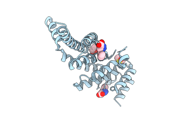 |
Organism: Homo sapiens
Method: X-RAY DIFFRACTION Release Date: 2025-12-03 Classification: PROTEIN BINDING Ligands: GOL, UJK, DMS |
 |
Organism: Homo sapiens
Method: X-RAY DIFFRACTION Release Date: 2025-12-03 Classification: PROTEIN BINDING Ligands: UUG, DMS |
 |
Organism: Rabbit hemorrhagic disease virus
Method: ELECTRON MICROSCOPY Release Date: 2025-12-03 Classification: VIRAL PROTEIN |
 |
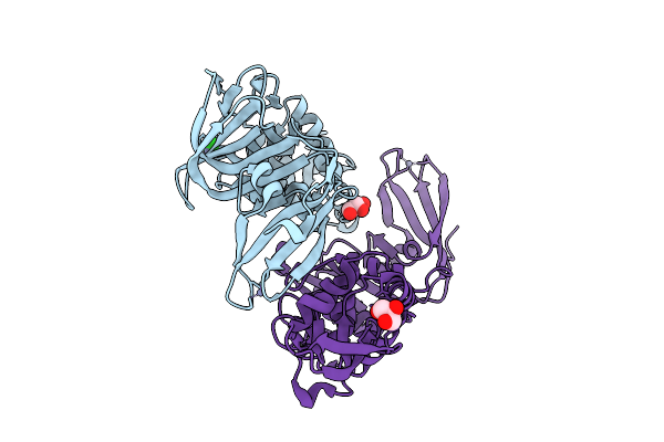
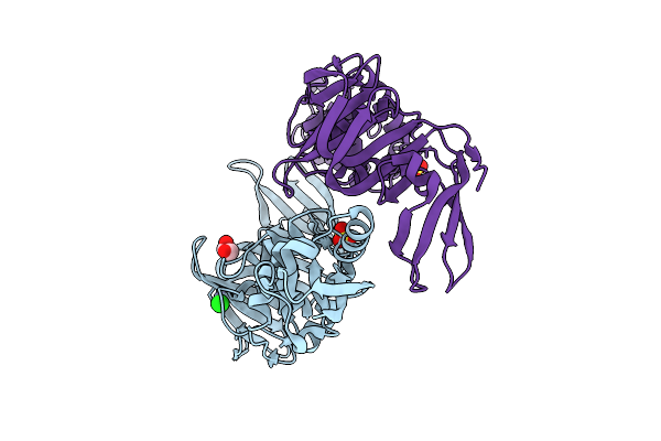
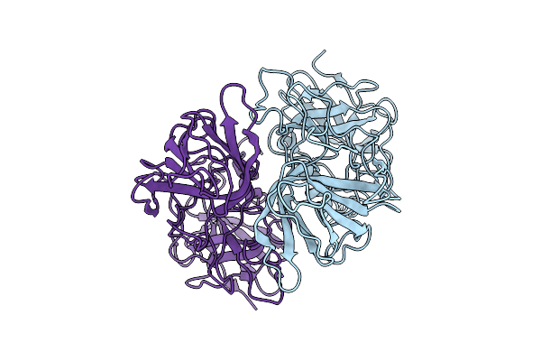
Crystal Structure Of Zika Virus Ns2B-Ns3 Protease In Complex With Compound 2
Organism: Zika virus
Method: X-RAY DIFFRACTION Release Date: 2025-12-03 Classification: HYDROLASE Ligands: A1I16, ACY |
 |
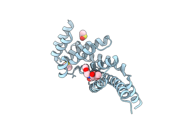
Organism: Homo sapiens
Method: X-RAY DIFFRACTION Release Date: 2025-12-03 Classification: CELL CYCLE Ligands: A1L6I |