
Search Count: 55
 |
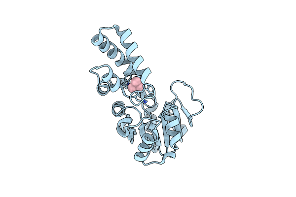
Organism: Staphylococcus aureus (strain mu3 / atcc 700698)
Method: X-RAY DIFFRACTION Resolution:2.19 Å Release Date: 2025-02-19 Classification: ANTIBIOTIC Ligands: A1EEE, MPD, TBU |
 |
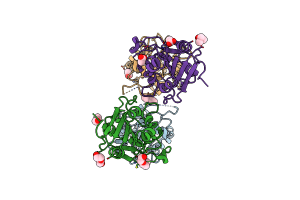
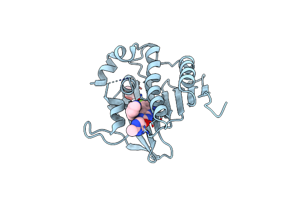
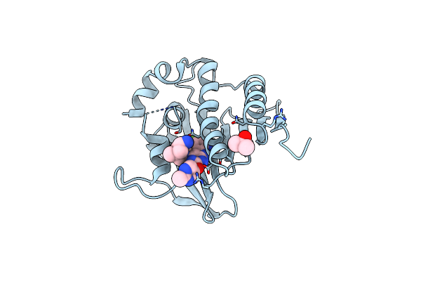
Flavin Mononucleotide-Dependent Nitroreductase B.Thetaiotaomicron (Bt_1680)
Organism: Bacteroides thetaiotaomicron
Method: X-RAY DIFFRACTION Resolution:1.80 Å Release Date: 2024-03-27 Classification: OXIDOREDUCTASE Ligands: CIT, TBU |
 |
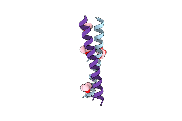
Structure Of A Cyclic Beta-Hairpin Peptide Derived From Neuronal Nitric Oxide Synthase (T112W/T116E Variant)
Organism: Homo sapiens
Method: X-RAY DIFFRACTION Resolution:1.44 Å Release Date: 2022-12-21 Classification: PROTEIN BINDING Ligands: TBU |
 |
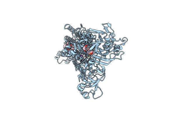
Organism: Bdellovibrio bacteriovorus (strain atcc 15356 / dsm 50701 / ncib 9529 / hd100)
Method: X-RAY DIFFRACTION Resolution:1.50 Å Release Date: 2021-05-05 Classification: UNKNOWN FUNCTION Ligands: MPD, GOL, TBU |
 |
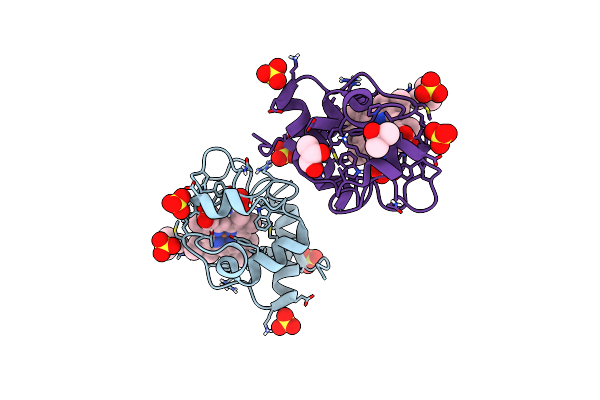
Organism: Homo sapiens
Method: X-RAY DIFFRACTION Resolution:2.25 Å Release Date: 2020-12-23 Classification: OXIDOREDUCTASE Ligands: OGA, FE2, TBU, GLY, CA, CL |
 |
Crystal Structure Of Human Methionine Aminopeptidase-2 In Complex With An Inhibitor Thiophene-2-Sulfonic Acid (4-Fluoro-Benzyl)-(4H-[1,2,4]Triazol-3-Ylmethyl)-Amide
Organism: Homo sapiens
Method: X-RAY DIFFRACTION Resolution:1.62 Å Release Date: 2019-05-01 Classification: HYDROLASE Ligands: EDO, HZT, TBU, MN |
 |
Organism: Homo sapiens
Method: X-RAY DIFFRACTION Resolution:1.82 Å Release Date: 2018-10-17 Classification: OXIDOREDUCTASE Ligands: FE, ZN, AKG, TBU |
 |
Organism: Homo sapiens
Method: X-RAY DIFFRACTION Resolution:1.97 Å Release Date: 2018-07-04 Classification: HYDROLASE Ligands: AR6, ZN, TBU, GOL |
 |
Organism: Homo sapiens, Synthetic construct
Method: X-RAY DIFFRACTION Resolution:2.10 Å Release Date: 2018-01-17 Classification: Hydrolase/Inhibitor Ligands: TBU, CA |
 |
The Structural Basis Of The Histone Demethylase Kdm6B Histone 3 Lysine 27 Specificity
Organism: Homo sapiens
Method: X-RAY DIFFRACTION Resolution:2.14 Å Release Date: 2017-09-20 Classification: OXIDOREDUCTASE Ligands: FE, ZN, AKG, EDO, TBU |
 |
Organism: Rattus norvegicus
Method: X-RAY DIFFRACTION Resolution:1.70 Å Release Date: 2017-09-20 Classification: PROTEIN BINDING Ligands: CA, TBU, ACT |
 |
Structure Of Human Methionine Aminopeptidase 2 With Covalent Spiroepoxytriazole Inhibitor (-)-31B
Organism: Homo sapiens
Method: X-RAY DIFFRACTION Resolution:1.49 Å Release Date: 2016-01-13 Classification: HYDROLASE Ligands: 94A, CO, TBU |
 |
Structure Of Human Methionine Aminopeptidase-2 Complexed With Spiroepoxytriazole Inhibitor (+)-31B
Organism: Homo sapiens
Method: X-RAY DIFFRACTION Release Date: 2016-01-13 Classification: HYDROLASE Ligands: 57R, CO, TBU, EDO |
 |
Organism: Thermococcus onnurineus (strain na1)
Method: X-RAY DIFFRACTION Resolution:2.00 Å Release Date: 2015-04-22 Classification: HYDROLASE Ligands: TBU |
 |
Crystal Structure Of Ahp1 From Saccharomyces Cerevisiae. Investigating The Electron Transfers.
Organism: Saccharomyces cerevisiae
Method: X-RAY DIFFRACTION Resolution:2.20 Å Release Date: 2015-02-04 Classification: OXIDOREDUCTASE Ligands: PEG, ACY, TBU |
 |
Organism: Oryctolagus cuniculus
Method: X-RAY DIFFRACTION Resolution:2.50 Å Release Date: 2014-10-01 Classification: HYDROLASE Ligands: TG1, GOL, K, MG, TBU, SO4 |
 |
Crystal Structure Of An Oxidized Form Of Yeast Iso-1-Cytochrome C At Ph 8.8
Organism: Saccharomyces cerevisiae
Method: X-RAY DIFFRACTION Resolution:1.45 Å Release Date: 2014-06-04 Classification: ELECTRON TRANSPORT Ligands: HEC, SO4, TBU, GOL |
 |
Organism: Synthetic construct
Method: X-RAY DIFFRACTION Resolution:1.88 Å Release Date: 2014-02-05 Classification: DE NOVO PROTEIN Ligands: TBU, GOL |
 |
The Dna Gyrase B Atp Binding Domain Of Enterococcus Faecalis In Complex With A Small Molecule Inhibitor
Organism: Enterococcus faecalis
Method: X-RAY DIFFRACTION Resolution:1.30 Å Release Date: 2014-01-15 Classification: ISOMERASE/ISOMERASE INHIBITOR Ligands: DOO, TBU |
 |
Dna Gyrase Atp Binding Domain Of Enterococcus Faecalis In Complex With A Small Molecule Inhibitor (4-[(1S,5R,6R)-6-Amino-1-Methyl-3-Azabicyclo[3.2.0]Hept-3-Yl]-6-Fluoro-N-Methyl-2-[(2-Methylpyrimidin-5-Yl)Oxy]-9H-Pyrimido[4,5-B]Indol-8-Amine)
Organism: Enterococcus faecalis
Method: X-RAY DIFFRACTION Resolution:1.75 Å Release Date: 2014-01-15 Classification: Isomerase/isomerase inhibitor Ligands: 920, TBU |