
Search Count: 7
 |
Crystal Structure Of Sialic Acid Binding Protein From Haemophilus Ducreyi With Neu5Gc
Organism: Haemophilus ducreyi 35000hp
Method: X-RAY DIFFRACTION Resolution:2.48 Å Release Date: 2018-10-24 Classification: SUGAR BINDING PROTEIN Ligands: NGE |
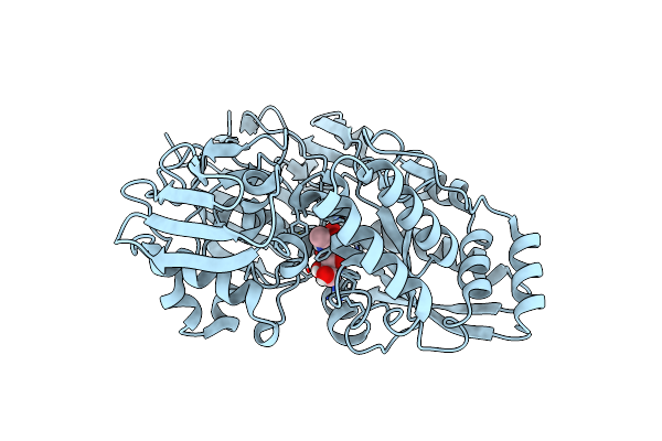 |
Crystal Structure Of Sialic Acid Binding Protein From Haemophilus Ducreyi With Neu5Ac
Organism: Haemophilus ducreyi 35000hp
Method: X-RAY DIFFRACTION Resolution:1.49 Å Release Date: 2018-10-24 Classification: SUGAR BINDING PROTEIN Ligands: SLB |
 |
Organism: Haemophilus ducreyi
Method: X-RAY DIFFRACTION Resolution:2.19 Å Release Date: 2018-10-24 Classification: SUGAR BINDING PROTEIN |
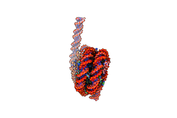 |
Crystal Structure Of A 197-Bp Palindromic 601L Nucleosome In Complex With Linker Histone H1
Organism: Xenopus laevis, Synthetic construct
Method: X-RAY DIFFRACTION Resolution:5.40 Å Release Date: 2017-05-17 Classification: CHROMATIN BINDING PROTEIN / DNA |
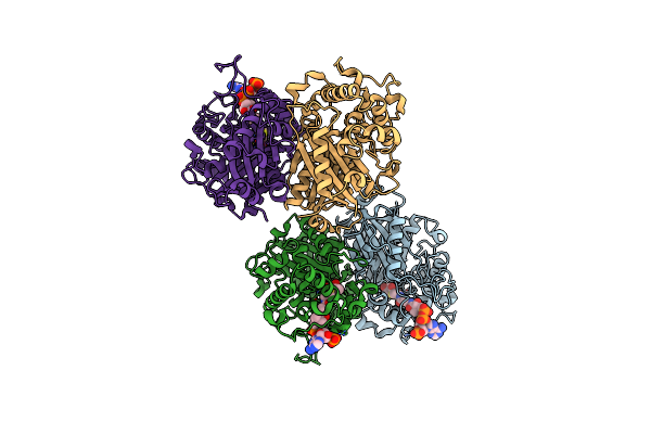 |
Crystal Structure Of Scp2 Thiolase From Leishmania Mexicana. Complex Of The C123A Mutant With Acetoacetyl-Coa.
Organism: Leishmania mexicana
Method: X-RAY DIFFRACTION Resolution:1.98 Å Release Date: 2017-01-18 Classification: TRANSFERASE Ligands: CAA |
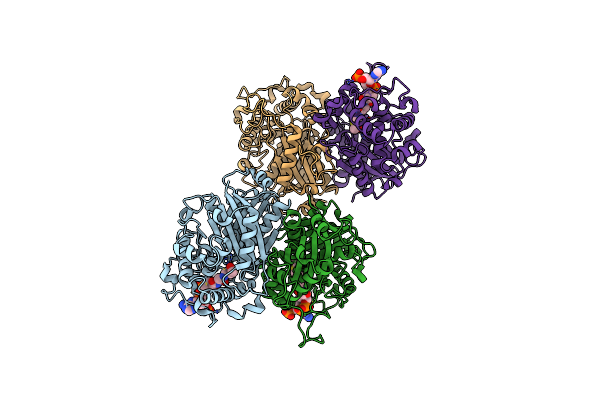 |
Crystal Structure Of Scp2 Thiolase From Leishmania Mexicana. Complex Of The C123A Mutant With Acetyl-Coa.
Organism: Leishmania mexicana (strain mhom/gt/2001/u1103)
Method: X-RAY DIFFRACTION Resolution:2.25 Å Release Date: 2017-01-18 Classification: TRANSFERASE Ligands: ACO |
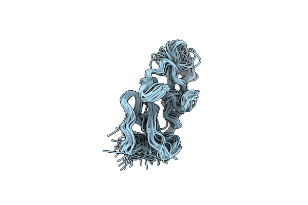 |
Solution Structure Of An Egf Module Pair From The Plasmodium Falciparum Merozoite Surface Protein 1
Organism: Plasmodium falciparum
Method: SOLUTION NMR Release Date: 1999-05-28 Classification: SURFACE PROTEIN |