
Search Count: 12
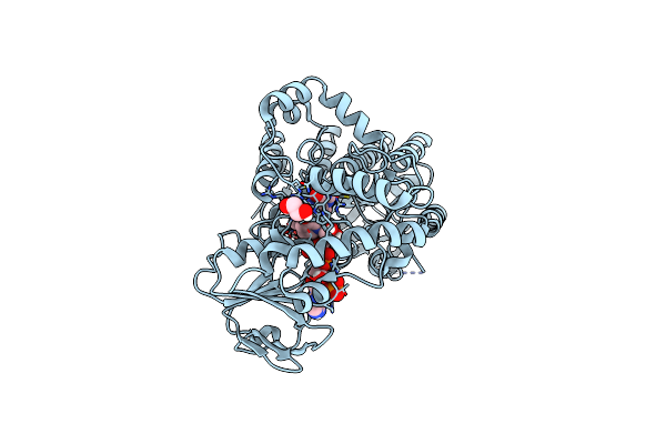 |
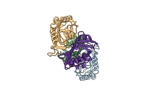
Crystal Structure Of Phenylacetone Monooxygenase Mutant Pm1 Bound To Fad And Nadp
Organism: Thermobifida fusca yx
Method: X-RAY DIFFRACTION Resolution:2.00 Å Release Date: 2024-12-18 Classification: OXIDOREDUCTASE Ligands: NAP, FAD, EDO |
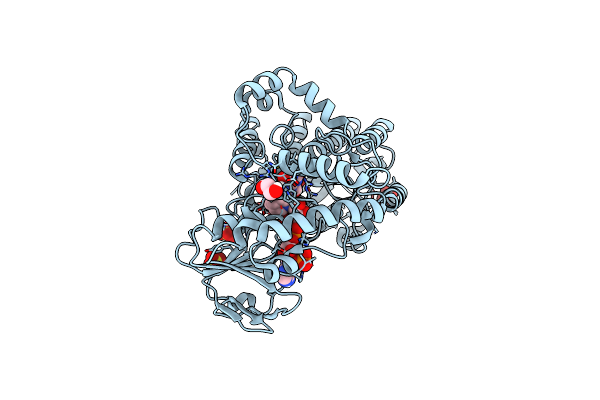 |
Crystal Structure Of Phenylacetone Monooxygenase Mutant Pm2 Bound To Fad And Nadp
Organism: Thermobifida fusca yx
Method: X-RAY DIFFRACTION Resolution:2.13 Å Release Date: 2024-12-18 Classification: OXIDOREDUCTASE Ligands: FAD, NAP, EDO, SO4 |
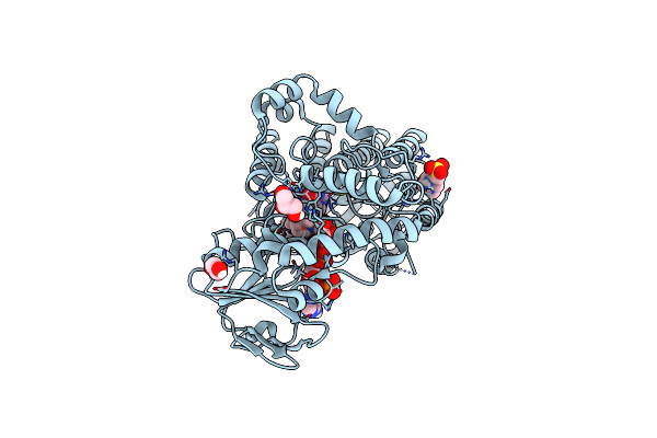 |
Crystal Structure Of Phenylacetone Monooxygenase Mutant Pm3 Bound To Fad And Nadp
Organism: Thermobifida fusca yx
Method: X-RAY DIFFRACTION Resolution:2.00 Å Release Date: 2024-12-18 Classification: OXIDOREDUCTASE Ligands: FAD, NAP, MES, EDO, GOL |
 |
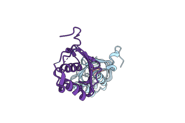
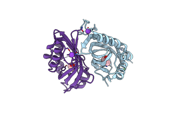
Crystal Structure Of Alcohol Dehydrogenase M5 From Burkholderia Gladioli With Nadp
Organism: Burkholderia gladioli bsr3
Method: X-RAY DIFFRACTION Resolution:2.27 Å Release Date: 2024-03-06 Classification: OXIDOREDUCTASE Ligands: NAP |
 |
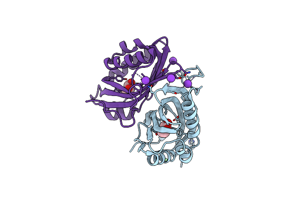
Crystal Structure Of Alcohol Dehydrogenase From Burkholderia Gladioli With Nadp
Organism: Burkholderia gladioli (strain bsr3)
Method: X-RAY DIFFRACTION Resolution:2.28 Å Release Date: 2024-02-28 Classification: OXIDOREDUCTASE Ligands: NAP |
 |
Organism: Burkholderia gladioli (strain bsr3)
Method: X-RAY DIFFRACTION Resolution:2.54 Å Release Date: 2024-02-28 Classification: OXIDOREDUCTASE |
 |
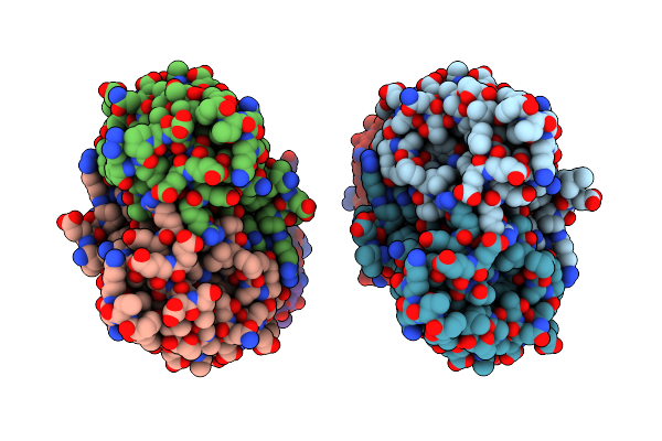
Crystal Structure Of Alcohol Dehydrogenase M4 Mutant From Burkholderia Gladioli
Organism: Burkholderia gladioli (strain bsr3)
Method: X-RAY DIFFRACTION Resolution:2.38 Å Release Date: 2024-02-28 Classification: OXIDOREDUCTASE |
 |
Organism: Rhodococcus erythropolis
Method: X-RAY DIFFRACTION Resolution:2.01 Å Release Date: 2017-07-12 Classification: HYDROLASE Ligands: 6VV |
 |
Organism: Rhodococcus erythropolis
Method: X-RAY DIFFRACTION Resolution:2.50 Å Release Date: 2016-07-20 Classification: HYDROLASE Ligands: 3ZS, K, NI |
 |
Organism: Rhodococcus erythropolis
Method: X-RAY DIFFRACTION Resolution:2.70 Å Release Date: 2016-07-20 Classification: HYDROLASE Ligands: NI, 3ZQ, K, CL |
 |
Organism: Rhodococcus erythropolis
Method: X-RAY DIFFRACTION Resolution:2.24 Å Release Date: 2016-07-13 Classification: HYDROLASE |
 |
Organism: Rhodococcus erythropolis
Method: X-RAY DIFFRACTION Resolution:3.00 Å Release Date: 2016-07-13 Classification: HYDROLASE |