
Search Count: 353
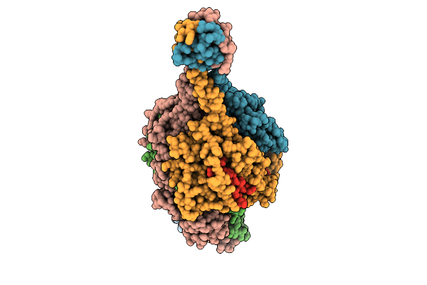 |
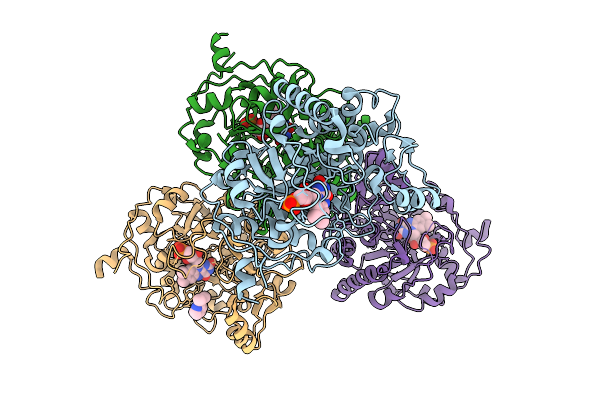
Respiratory Syncytial Virus Pre-F Trimer Bound By Neutralizing Antibody Pr306007
Organism: Respiratory syncytial virus a2, Homo sapiens
Method: ELECTRON MICROSCOPY Release Date: 2025-11-12 Classification: STRUCTURAL PROTEIN/IMMUNE SYSTEM |
 |
Crystal Structure Of A Coronaviral M Protein In Complex With A C-Terminal Peptide Of The N Protein
Organism: Pipistrellus bat coronavirus hku5
Method: X-RAY DIFFRACTION Release Date: 2025-11-05 Classification: MEMBRANE PROTEIN Ligands: N8E |
 |
Organism: Homo sapiens
Method: ELECTRON MICROSCOPY Release Date: 2025-11-05 Classification: HYDROLASE |
 |
Crystal Structure Of The Carbamoyl N-Methyltransferase Asc-Orf2 Complexed With Sah
Organism: Amycolatopsis alba dsm 44262
Method: X-RAY DIFFRACTION Release Date: 2025-10-29 Classification: BIOSYNTHETIC PROTEIN Ligands: SAH |
 |
Organism: Sordaria araneosa
Method: X-RAY DIFFRACTION Release Date: 2025-10-22 Classification: BIOSYNTHETIC PROTEIN Ligands: A1LYT, MPD |
 |
The Structure Of Sdng Covalently Binding With The Cope Rearrangement Product
Organism: Sordaria araneosa
Method: X-RAY DIFFRACTION Release Date: 2025-10-22 Classification: BIOSYNTHETIC PROTEIN Ligands: MPD, A1LZG |
 |
Organism: Sordaria araneosa
Method: X-RAY DIFFRACTION Release Date: 2025-10-22 Classification: BIOSYNTHETIC PROTEIN Ligands: A1LZH, A1LZK |
 |
Organism: Sordaria araneosa
Method: X-RAY DIFFRACTION Release Date: 2025-10-22 Classification: BIOSYNTHETIC PROTEIN Ligands: MPD |
 |
Organism: Sordaria araneosa
Method: X-RAY DIFFRACTION Release Date: 2025-10-22 Classification: BIOSYNTHETIC PROTEIN Ligands: A1LYT |
 |
Organism: Homo sapiens, Vicugna pacos
Method: X-RAY DIFFRACTION Release Date: 2025-10-01 Classification: IMMUNE SYSTEM Ligands: EDO, SO4 |
 |
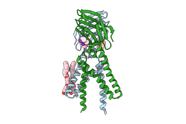
The Glycoprotein E Of Varicella-Zoster Virus In Complex With Wll-1/Wll-28 Fab
Organism: Varicella-zoster virus (strain oka vaccine), Homo sapiens
Method: ELECTRON MICROSCOPY Release Date: 2025-09-03 Classification: VIRAL PROTEIN/IMMUNE SYSTEM |
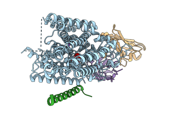 |
Structure Of Hsglt2-Map17 Complex In The Substrate-Bound Occluded Conformation
Organism: Homo sapiens, Mus musculus
Method: ELECTRON MICROSCOPY Release Date: 2025-09-03 Classification: TRANSPORT PROTEIN |
 |
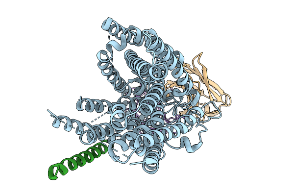
Structure Of Hsglt2-Map17 Complex In The Substrate-Free Inward-Facing Conformation In The Presence Of Sodium
Organism: Homo sapiens, Mus musculus
Method: ELECTRON MICROSCOPY Release Date: 2025-09-03 Classification: TRANSPORT PROTEIN |
 |
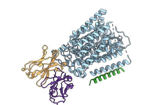
Structure Of Hsglt2-Map17 Complex In The Substrate-Free Inward-Facing Conformation In The Presence Of Potassium
Organism: Homo sapiens, Mus musculus
Method: ELECTRON MICROSCOPY Release Date: 2025-09-03 Classification: TRANSPORT PROTEIN |
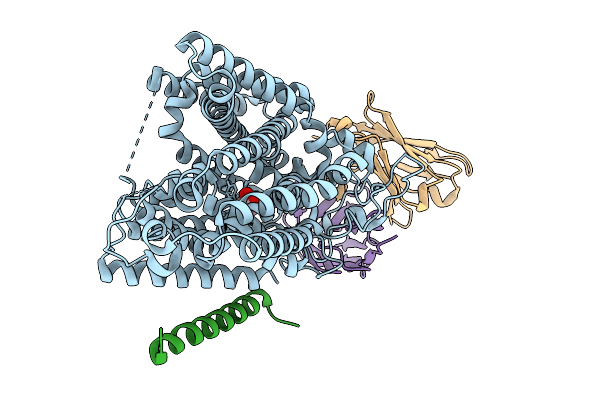 |
Structure Of Hsglt2-Map17 Complex In The Substrate-Bound Inward-Facing Conformation
Organism: Homo sapiens, Mus musculus
Method: ELECTRON MICROSCOPY Release Date: 2025-09-03 Classification: TRANSPORT PROTEIN Ligands: A1EFW |
 |
Organism: Saccharomyces cerevisiae s288c
Method: X-RAY DIFFRACTION Release Date: 2025-09-03 Classification: OXIDOREDUCTASE Ligands: FMN, A1EG5 |
 |
Organism: Henipavirus nipahense, Mus musculus
Method: ELECTRON MICROSCOPY Release Date: 2025-07-02 Classification: VIRAL PROTEIN/IMMUNE SYSTEM |
 |
Organism: Penicillium
Method: X-RAY DIFFRACTION Release Date: 2025-06-18 Classification: BIOSYNTHETIC PROTEIN Ligands: FE |
 |
Organism: Mus musculus
Method: ELECTRON MICROSCOPY Release Date: 2025-04-23 Classification: PROTEIN FIBRIL |
 |
Organism: Mus musculus
Method: ELECTRON MICROSCOPY Release Date: 2025-04-23 Classification: PROTEIN FIBRIL |