
Search Count: 331
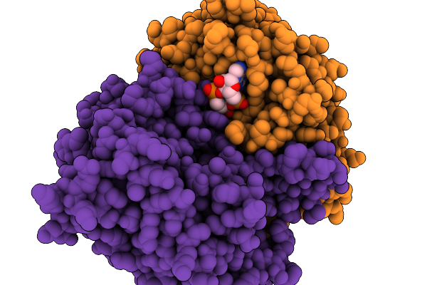 |
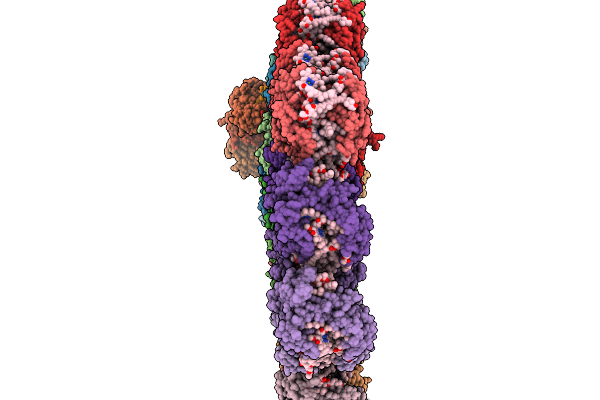
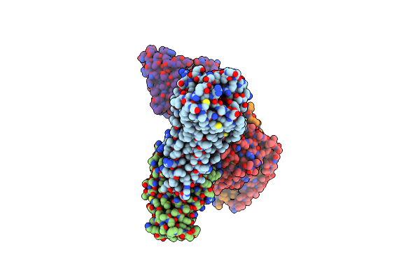
Organism: Acinetobacter baumannii
Method: ELECTRON MICROSCOPY Release Date: 2025-11-19 Classification: HYDROLASE Ligands: 4BW |
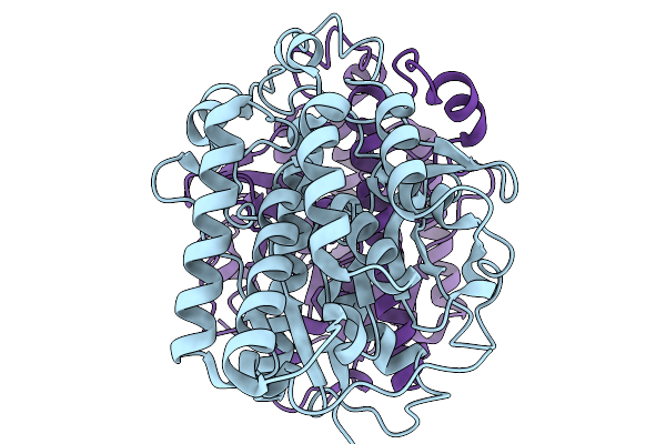 |
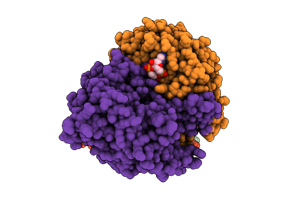
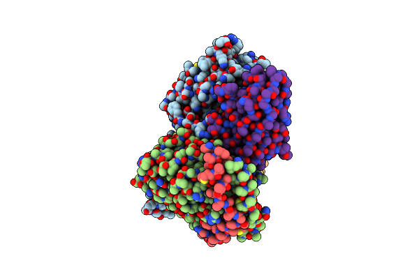
Organism: Acinetobacter baumannii
Method: ELECTRON MICROSCOPY Release Date: 2025-11-19 Classification: HYDROLASE |
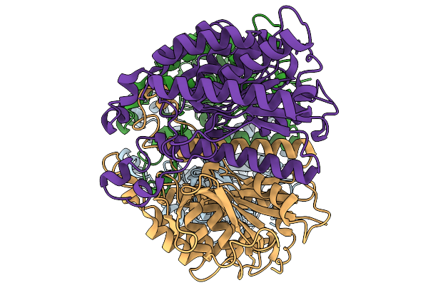 |
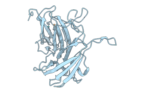
Organism: Acinetobacter baumannii
Method: ELECTRON MICROSCOPY Release Date: 2025-11-19 Classification: HYDROLASE |
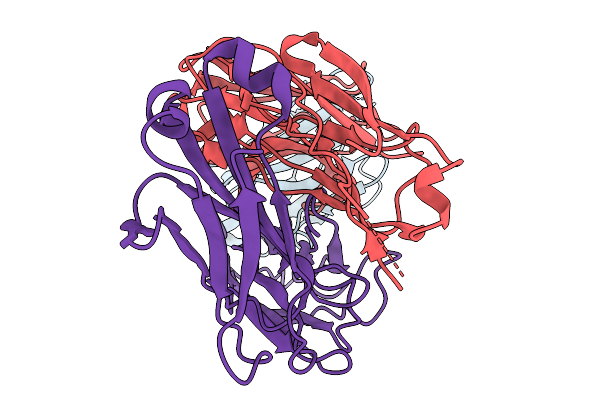 |
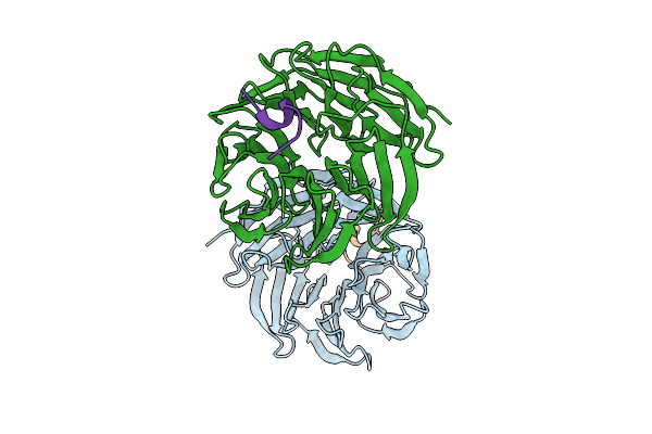
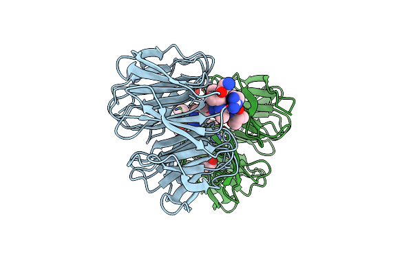
Structure Of Nectin-4 D1 Domain In Complex With The Fab Fragment Of 9Mw2821 Mab
Organism: Mus musculus, Homo sapiens
Method: ELECTRON MICROSCOPY Release Date: 2025-11-19 Classification: IMMUNE SYSTEM |
 |
Organism: Emiliania huxleyi
Method: ELECTRON MICROSCOPY Release Date: 2025-11-19 Classification: PHOTOSYNTHESIS Ligands: CLA, KC2, DD6, LMG, A86, LHG, SQD, A1EB1, A1EB4, BCR, PQN, SF4, DGD |
 |
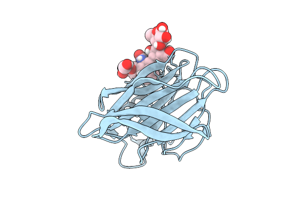
Organism: Acinetobacter baumannii
Method: ELECTRON MICROSCOPY Release Date: 2025-11-12 Classification: HYDROLASE Ligands: 4BW |
 |
Organism: Homo sapiens
Method: X-RAY DIFFRACTION Release Date: 2025-11-05 Classification: IMMUNE SYSTEM |
 |
Organism: Homo sapiens
Method: X-RAY DIFFRACTION Release Date: 2025-11-05 Classification: IMMUNE SYSTEM Ligands: PO4 |
 |
Organism: Streptomyces coelicolor a3(2)
Method: X-RAY DIFFRACTION Release Date: 2025-10-01 Classification: OXIDOREDUCTASE Ligands: CU, RU |
 |
Organism: Mus musculus
Method: ELECTRON MICROSCOPY Release Date: 2025-08-27 Classification: MEMBRANE PROTEIN |
 |
Organism: Mus musculus
Method: ELECTRON MICROSCOPY Release Date: 2025-08-27 Classification: MEMBRANE PROTEIN |
 |
Organism: Homo sapiens, Mus musculus
Method: X-RAY DIFFRACTION Release Date: 2025-08-13 Classification: NUCLEAR PROTEIN |
 |
Organism: Coronaviridae
Method: ELECTRON MICROSCOPY Release Date: 2025-07-16 Classification: VIRAL PROTEIN Ligands: NAG |
 |
Organism: Rotavirus
Method: X-RAY DIFFRACTION Release Date: 2025-07-09 Classification: VIRAL PROTEIN |
 |
Organism: Rotavirus
Method: X-RAY DIFFRACTION Release Date: 2025-07-09 Classification: VIRAL PROTEIN |
 |
Organism: Homo sapiens, Vicugna pacos
Method: ELECTRON MICROSCOPY Release Date: 2025-06-11 Classification: MEMBRANE PROTEIN Ligands: UTP |
 |
Organism: Homo sapiens, Vicugna pacos
Method: ELECTRON MICROSCOPY Release Date: 2025-06-11 Classification: MEMBRANE PROTEIN Ligands: ATP |
 |
Organism: Homo sapiens, Mus musculus
Method: ELECTRON MICROSCOPY Release Date: 2025-06-11 Classification: MEMBRANE PROTEIN Ligands: ATP |
 |
Organism: Homo sapiens, Vicugna pacos
Method: ELECTRON MICROSCOPY Release Date: 2025-06-11 Classification: MEMBRANE PROTEIN |
 |
Organism: Homo sapiens
Method: X-RAY DIFFRACTION Resolution:1.85 Å Release Date: 2025-05-21 Classification: CYTOSOLIC PROTEIN Ligands: EDO |