
Search Count: 659
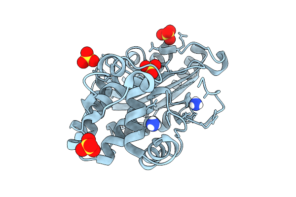 |
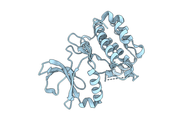
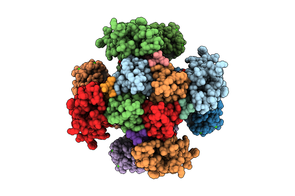
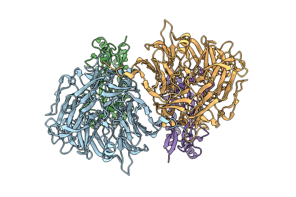
Structure Of P167S/D240G/D172A/S104G Blac From Mycobacterium Tuberculosis At Ph 5.5
Organism: Mycobacterium tuberculosis
Method: X-RAY DIFFRACTION Release Date: 2025-10-22 Classification: HYDROLASE Ligands: NH4, SO4 |
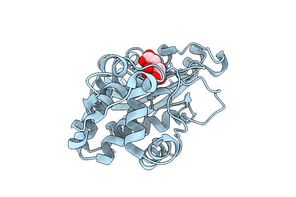 |
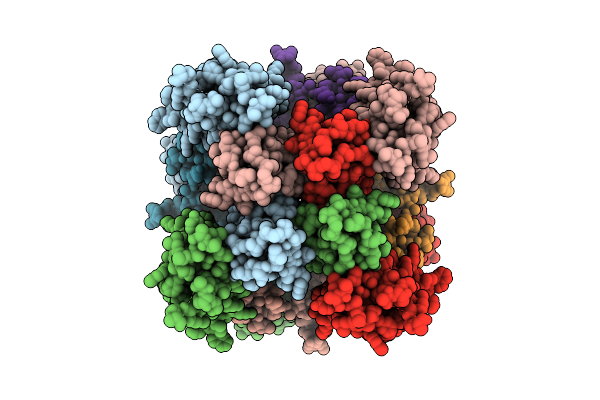
Organism: Mycobacterium tuberculosis
Method: X-RAY DIFFRACTION Release Date: 2025-10-22 Classification: HYDROLASE Ligands: CIT |
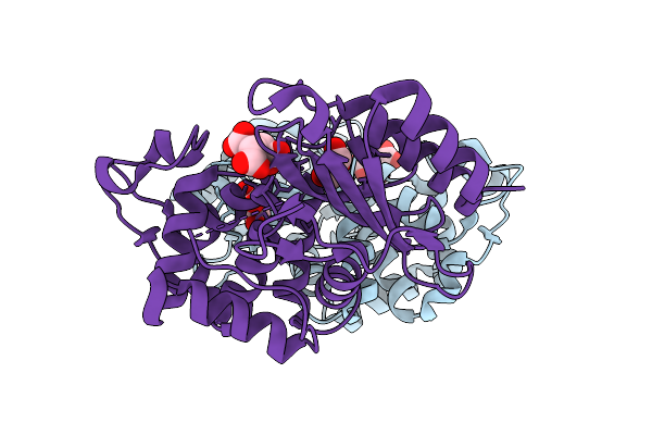 |
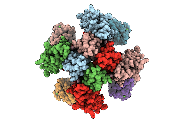
Structure Of P167S/D240G/D172A/S104G/H184R Blac From Mycobacterium Tuberculosis
Organism: Mycobacterium tuberculosis
Method: X-RAY DIFFRACTION Release Date: 2025-10-22 Classification: HYDROLASE Ligands: FLC, GOL |
 |
Organism: Pseudomonadota bacterium
Method: X-RAY DIFFRACTION Release Date: 2025-10-22 Classification: HYDROLASE Ligands: GOL |
 |
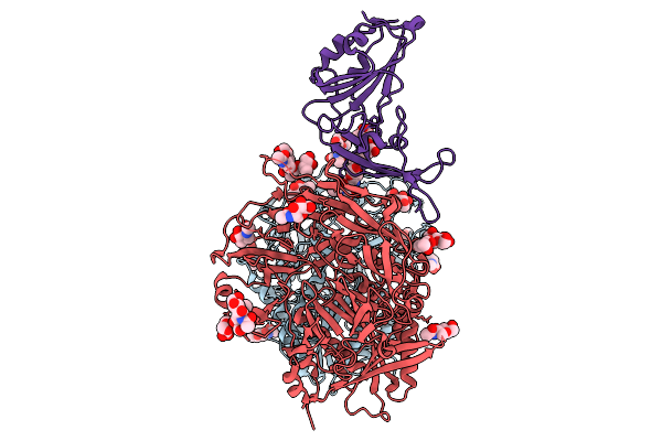 |
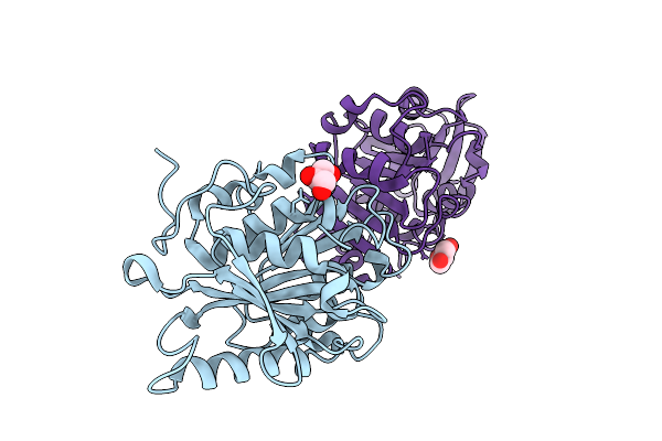
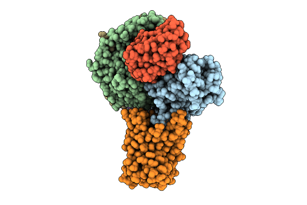
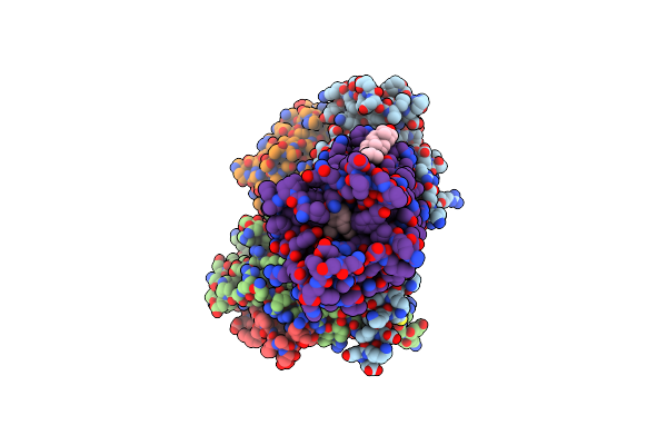
Cryo-Em Structure Of Mjhku4R-Cov-1 Receptor-Binding Domain Complexed With Human Cd26
Organism: Homo sapiens, Tylonycteris bat coronavirus hku4
Method: ELECTRON MICROSCOPY Release Date: 2025-09-03 Classification: VIRAL PROTEIN/HYDROLASE Ligands: NAG |
 |
Organism: Homo sapiens
Method: ELECTRON MICROSCOPY Release Date: 2025-09-03 Classification: MEMBRANE PROTEIN Ligands: CA, A1BBG |
 |
Organism: Homo sapiens
Method: ELECTRON MICROSCOPY Release Date: 2025-08-20 Classification: MEMBRANE PROTEIN Ligands: CA |
 |
Organism: Homo sapiens
Method: ELECTRON MICROSCOPY Release Date: 2025-08-20 Classification: MEMBRANE PROTEIN Ligands: PT5, CA |
 |
Organism: Homo sapiens
Method: ELECTRON MICROSCOPY Release Date: 2025-08-20 Classification: MEMBRANE PROTEIN Ligands: CA, PT5 |
 |
Organism: Homo sapiens
Method: ELECTRON MICROSCOPY Release Date: 2025-08-20 Classification: MEMBRANE PROTEIN Ligands: PT5, CA |
 |
Organism: Spodoptera
Method: ELECTRON MICROSCOPY Release Date: 2025-08-13 Classification: MEMBRANE PROTEIN Ligands: AND |
 |
Organism: Homo sapiens
Method: ELECTRON MICROSCOPY Release Date: 2025-08-13 Classification: MEMBRANE PROTEIN Ligands: A1EJ7 |
 |
Organism: Homo sapiens
Method: ELECTRON MICROSCOPY Release Date: 2025-07-30 Classification: MEMBRANE PROTEIN |
 |
Organism: Methylorubrum extorquens
Method: ELECTRON MICROSCOPY Release Date: 2025-07-23 Classification: PROTEIN BINDING |
 |
Organism: Methylorubrum extorquens
Method: ELECTRON MICROSCOPY Release Date: 2025-07-23 Classification: PROTEIN BINDING Ligands: PQQ |
 |
Organism: Homo sapiens, Lama glama, Escherichia coli
Method: ELECTRON MICROSCOPY Release Date: 2025-07-02 Classification: MEMBRANE PROTEIN Ligands: CLR, A1ESD |
 |
Organism: Homo sapiens, Lama glama, Escherichia coli, Mus musculus
Method: ELECTRON MICROSCOPY Release Date: 2025-07-02 Classification: MEMBRANE PROTEIN/IMMUNE SYSTEM Ligands: A1EQV |
 |
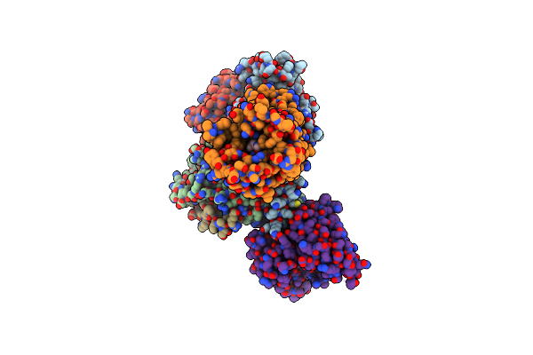
The Structure Of Smarcad1 Bound To The Hexasome In The Presence Of Adp-Befx
Organism: Homo sapiens, Xenopus laevis, Synthetic construct
Method: ELECTRON MICROSCOPY Release Date: 2025-04-30 Classification: DNA BINDING PROTEIN/DNA Ligands: ADP, BEF, MG |
 |
Organism: Homo sapiens, Lama glama
Method: ELECTRON MICROSCOPY Release Date: 2025-04-30 Classification: MEMBRANE PROTEIN Ligands: A1LXR |