
Search Count: 1,104
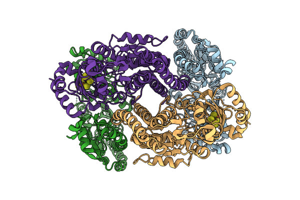 |
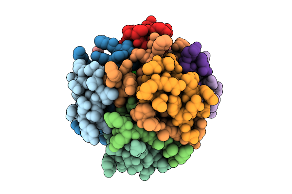
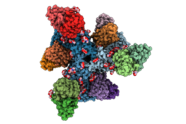
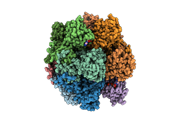
Rhodospirillum Rubrum Nitrogenase-Like Methylthio-Alkane Reductase Complex With An Oxidized P-Cluster
Organism: Rhodospirillum rubrum atcc 11170
Method: ELECTRON MICROSCOPY Release Date: 2025-11-05 Classification: OXIDOREDUCTASE Ligands: CLF |
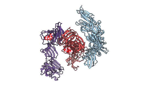 |
Crystal Structure Of Ha3 From Clostridium Botulinum Type B With Alpha2,3-Sialyllactose
Organism: Clostridium botulinum b1 str. okra
Method: X-RAY DIFFRACTION Release Date: 2025-11-05 Classification: TOXIN |
 |
Crystal Structure Of Ha3 From Clostridium Botulinum Type B With Alpha2,6-Sialyllactose
Organism: Clostridium botulinum b1 str. okra
Method: X-RAY DIFFRACTION Release Date: 2025-11-05 Classification: TOXIN Ligands: SIA |
 |
Organism: Homo sapiens
Method: X-RAY DIFFRACTION Release Date: 2025-10-29 Classification: HORMONE Ligands: CRS, ZN, CL, CA |
 |
Organism: Homo sapiens
Method: X-RAY DIFFRACTION Release Date: 2025-10-29 Classification: HORMONE Ligands: IPH, ZN, CL, CA |
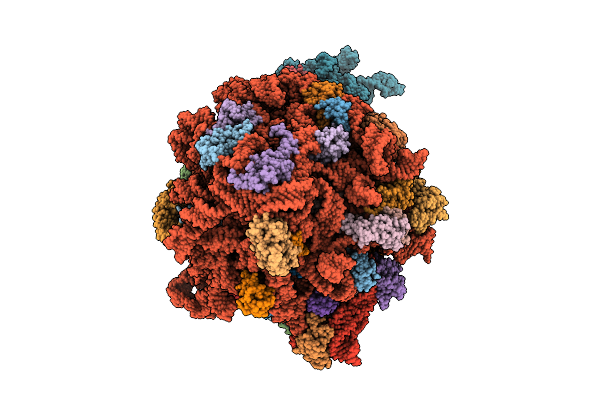 |
Organism: Escherichia coli
Method: ELECTRON MICROSCOPY Release Date: 2025-10-22 Classification: RIBOSOME Ligands: ZN, PRO |
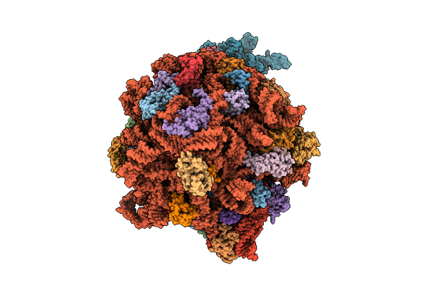 |
Structure Of E.Coli Ribosome In Complex With An Engineered Arrest Peptide And Trigger Factor
Organism: Escherichia coli
Method: ELECTRON MICROSCOPY Release Date: 2025-10-22 Classification: RIBOSOME Ligands: ZN, PRO |
 |
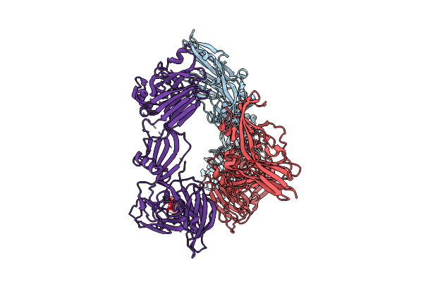
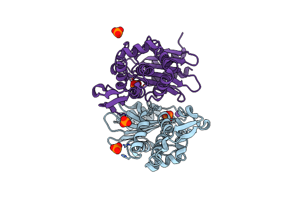
Crystal Structure Of Ospa St1 From B. Burgdorferi Bound To Monoclonal Antibody La-2
Organism: Mus musculus, Borreliella burgdorferi b31
Method: X-RAY DIFFRACTION Release Date: 2025-09-24 Classification: IMMUNE SYSTEM |
 |
Organism: Homo sapiens, Human immunodeficiency virus 1
Method: ELECTRON MICROSCOPY Release Date: 2025-07-30 Classification: VIRAL PROTEIN/Immune System Ligands: NAG |
 |
Bg505 Md39.3 Env Gp151 Mper Nanodisc In Complex With 10E8, Bg18 And Vrc01 Fabs (1X 10E8 Fab)
Organism: Homo sapiens, Human immunodeficiency virus 1
Method: ELECTRON MICROSCOPY Release Date: 2025-07-30 Classification: VIRAL PROTEIN/Immune System Ligands: NAG |
 |
Organism: Human parvovirus b19
Method: ELECTRON MICROSCOPY Release Date: 2025-07-09 Classification: DNA BINDING PROTEIN Ligands: ANP, MG |
 |
Organism: Human parvovirus b19
Method: ELECTRON MICROSCOPY Release Date: 2025-07-09 Classification: DNA BINDING PROTEIN/DNA Ligands: ANP, MG |
 |
Organism: Human parvovirus b19
Method: ELECTRON MICROSCOPY Release Date: 2025-07-09 Classification: DNA BINDING PROTEIN/DNA Ligands: ANP, MG |
 |
Organism: Human parvovirus b19
Method: ELECTRON MICROSCOPY Release Date: 2025-07-09 Classification: DNA BINDING PROTEIN Ligands: MG, ANP |
 |
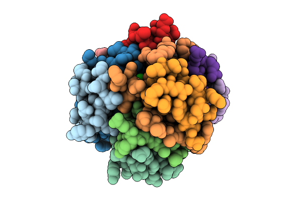
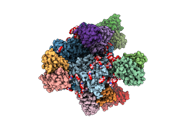
Organism: Saccharomyces cerevisiae
Method: X-RAY DIFFRACTION Release Date: 2025-06-25 Classification: RIBOSOME Ligands: OHX, MG, K, SPD, VDU, ZN, 5XU |
 |
Organism: Escherichia coli
Method: ELECTRON MICROSCOPY Release Date: 2025-06-18 Classification: RIBOSOME Ligands: ZN, PRO |
 |
Structure Of E.Coli Ribosome In Complex With An Engineered Arrest Peptide And Trigger Factor
Organism: Escherichia coli
Method: ELECTRON MICROSCOPY Release Date: 2025-06-18 Classification: RIBOSOME Ligands: ZN, PRO |
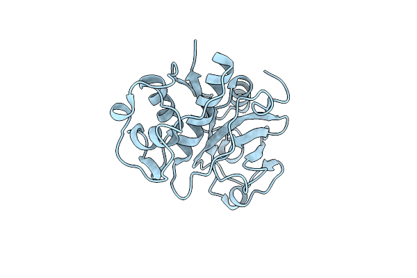 |
Organism: Carica papaya
Method: X-RAY DIFFRACTION Release Date: 2025-06-18 Classification: HYDROLASE |
 |
Organism: Escherichia coli
Method: X-RAY DIFFRACTION Release Date: 2025-06-18 Classification: HYDROLASE Ligands: PO4, K |
 |
X-Ray Diffraction Structure Of Ctx-M-14 Beta-Lactamase Co-Crystallized With Avibactam
Organism: Escherichia coli
Method: X-RAY DIFFRACTION Release Date: 2025-06-18 Classification: HYDROLASE Ligands: NXL, GOL, PO4 |