Planned Maintenance: Some services may turn out to be unavailable from 15th January, 2026 to 16th January, 2026. We apologize for the inconvenience!

Planned Maintenance: Some services may turn out to be unavailable from 15th January, 2026 to 16th January, 2026. We apologize for the inconvenience!
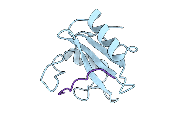 |
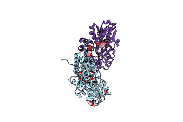
Structure Of The N-Sh2 Domain Of Shp2 In Complex With The Phosphoy627-Gab1 (613-651) Peptide
Organism: Homo sapiens
Method: X-RAY DIFFRACTION Release Date: 2025-12-31 Classification: SIGNALING PROTEIN |
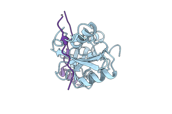 |
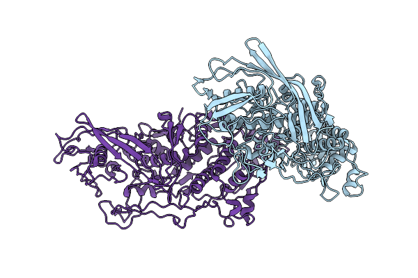
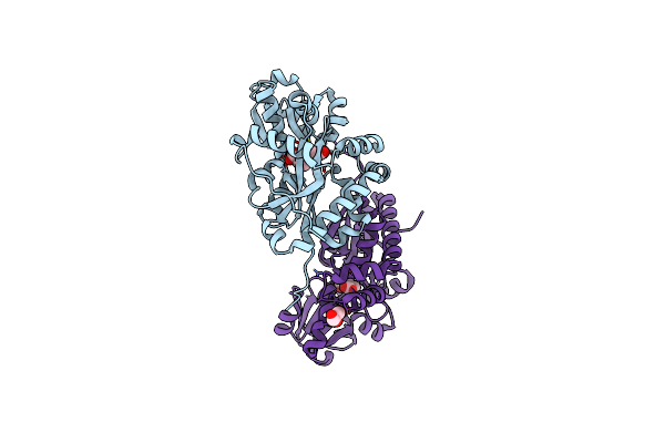
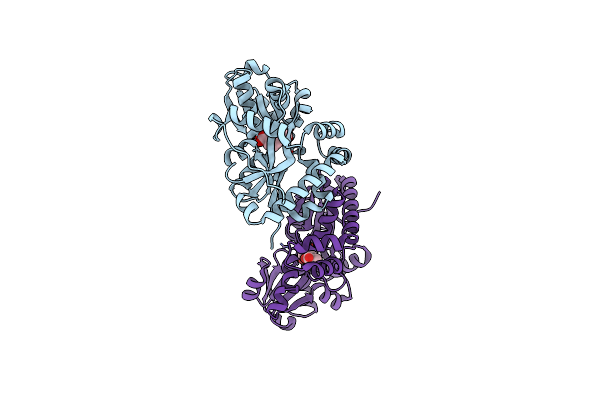
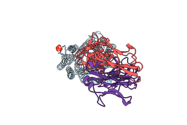
Micro-Ed Structure Of The Nsh2-Csh2 Tandem Domain Of Shp2 In Complex With The Bis-Phosphorylated Py627-Py659-Gab1 (613-694) Peptide
Organism: Homo sapiens
Method: ELECTRON CRYSTALLOGRAPHY Release Date: 2025-12-31 Classification: SIGNALING PROTEIN |
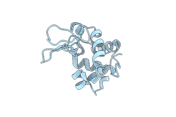 |
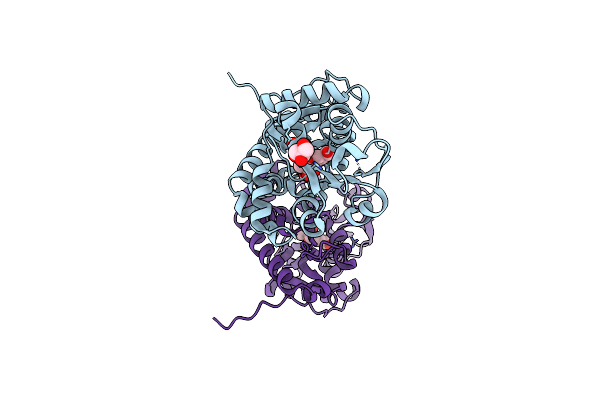
Hen Egg-White Lysozyme Structure Determined By 3Ded/Microed On A 200 Kev Microscope
Organism: Gallus gallus
Method: ELECTRON CRYSTALLOGRAPHY Resolution:2.80 Å Release Date: 2025-03-19 Classification: HYDROLASE |
 |
Organism: Saccharomyces cerevisiae
Method: ELECTRON MICROSCOPY Release Date: 2025-01-22 Classification: TRANSPORT PROTEIN Ligands: MG, GSP |
 |
Organism: Saccharomyces cerevisiae
Method: ELECTRON MICROSCOPY Release Date: 2025-01-22 Classification: TRANSPORT PROTEIN Ligands: MG, GSP |
 |
Organism: Saccharomyces cerevisiae
Method: ELECTRON MICROSCOPY Release Date: 2025-01-22 Classification: TRANSPORT PROTEIN Ligands: MG, GSP |
 |
Organism: Saccharomyces cerevisiae virus l-a
Method: ELECTRON MICROSCOPY Release Date: 2024-05-22 Classification: VIRUS |
 |
Organism: Saccharomyces cerevisiae
Method: ELECTRON MICROSCOPY Release Date: 2023-12-20 Classification: VIRUS |
 |
Organism: Streptomyces coelicolor
Method: X-RAY DIFFRACTION Resolution:1.92 Å Release Date: 2022-10-26 Classification: BIOSYNTHETIC PROTEIN Ligands: ISJ, GOL |
 |
Organism: Streptomyces coelicolor a3(2)
Method: X-RAY DIFFRACTION Resolution:1.91 Å Release Date: 2022-04-20 Classification: BIOSYNTHETIC PROTEIN Ligands: RDH |
 |
Organism: Streptomyces coelicolor a3(2)
Method: X-RAY DIFFRACTION Resolution:1.91 Å Release Date: 2022-04-20 Classification: BIOSYNTHETIC PROTEIN Ligands: ISJ, GOL |
 |
Organism: Streptomyces coelicolor a3(2)
Method: X-RAY DIFFRACTION Resolution:1.81 Å Release Date: 2022-04-20 Classification: BIOSYNTHETIC PROTEIN Ligands: GOL, RDH |
 |
Organism: Streptomyces coelicolor a3(2)
Method: X-RAY DIFFRACTION Resolution:2.01 Å Release Date: 2022-04-20 Classification: BIOSYNTHETIC PROTEIN Ligands: RDH, GOL |
 |
Organism: Streptomyces coelicolor a3(2)
Method: X-RAY DIFFRACTION Release Date: 2022-04-20 Classification: BIOSYNTHETIC PROTEIN Ligands: RDH |
 |
Organism: Streptomyces coelicolor (strain atcc baa-471 / a3(2) / m145)
Method: X-RAY DIFFRACTION Resolution:2.21 Å Release Date: 2021-10-06 Classification: BIOSYNTHETIC PROTEIN Ligands: GOL, RDH |
 |
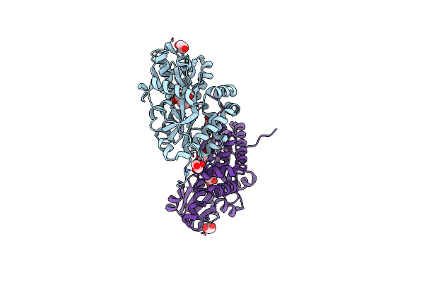
Molecular Structure Of Mouse Apoferritin Resolved At 2.7 Angstroms With The Glacios Cryo-Microscope
Organism: Mus musculus
Method: ELECTRON MICROSCOPY Resolution:2.73 Å Release Date: 2020-05-13 Classification: METAL BINDING PROTEIN Ligands: FE, MG |
 |
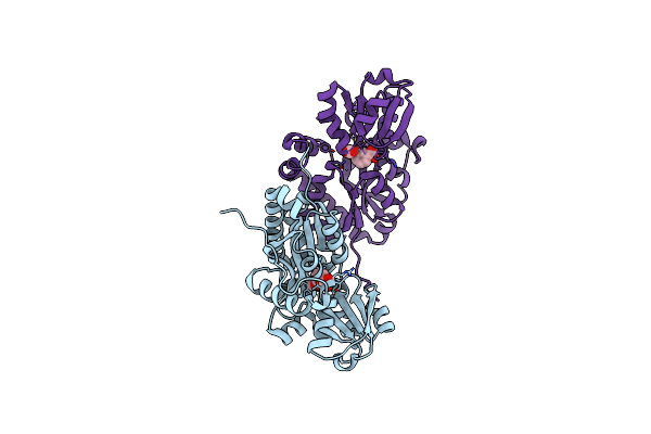
Organism: Porphyromonas gingivalis w83
Method: X-RAY DIFFRACTION Resolution:2.81 Å Release Date: 2019-03-06 Classification: TRANSFERASE Ligands: ZN |
 |
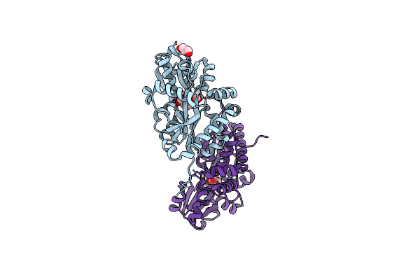
Organism: Tannerella forsythia (strain atcc 43037 / jcm 10827 / fdc 338)
Method: X-RAY DIFFRACTION Resolution:2.10 Å Release Date: 2019-03-06 Classification: TRANSFERASE Ligands: ZN, SO4 |
 |
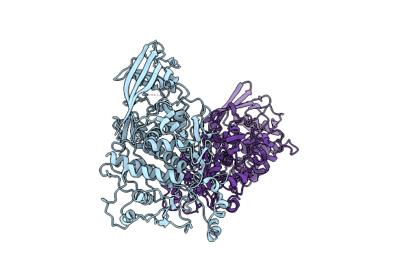
Crystal Structure Of E.Coli Multidrug/H+ Antiporter Mdfa In Outward Open Conformation With Bound Fab Fragment
Organism: Escherichia coli k-12, Mus musculus
Method: X-RAY DIFFRACTION Resolution:3.40 Å Release Date: 2018-10-03 Classification: TRANSPORT PROTEIN Ligands: SO4 |
 |
Structure Of Pyroglutamate-Abeta-Specific Fab C#17 In Complex With Human Abeta-Pe3-12Pegb
Organism: Mus musculus, Homo sapiens
Method: X-RAY DIFFRACTION Resolution:2.21 Å Release Date: 2017-06-28 Classification: IMMUNE SYSTEM |