
Search Count: 16
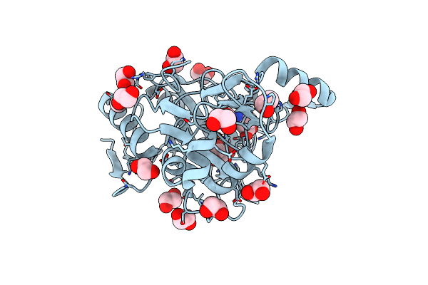 |
Organism: Streptomyces coeruleorubidus
Method: X-RAY DIFFRACTION Resolution:1.99 Å Release Date: 2022-03-23 Classification: BIOSYNTHETIC PROTEIN Ligands: FE2, 8S6, GOL, EDO |
 |
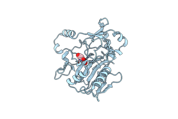
Organism: Streptomyces coeruleorubidus
Method: X-RAY DIFFRACTION Resolution:1.70 Å Release Date: 2021-01-06 Classification: OXIDOREDUCTASE Ligands: 7UC, ACT, FE2, CL |
 |
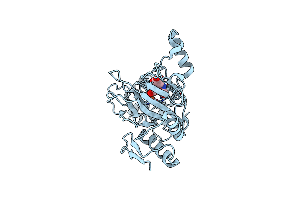
Organism: Streptomyces coeruleorubidus
Method: X-RAY DIFFRACTION Resolution:1.85 Å Release Date: 2021-01-06 Classification: OXIDOREDUCTASE Ligands: AKG, FE2, CL |
 |
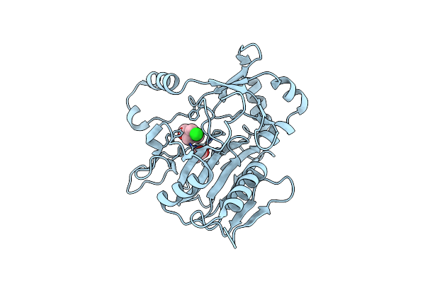
Organism: Streptomyces coeruleorubidus
Method: X-RAY DIFFRACTION Resolution:1.45 Å Release Date: 2021-01-06 Classification: OXIDOREDUCTASE Ligands: 7UC, AKG, ACT, FE |
 |
Scoe With Oxovanadium And The Caba Substrate Bound And His299 And Arg157 Flipped Out
Organism: Streptomyces coeruleorubidus
Method: X-RAY DIFFRACTION Resolution:2.10 Å Release Date: 2021-01-06 Classification: OXIDOREDUCTASE Ligands: 7UC, VVO, ACT, CL |
 |
Organism: Streptomyces coeruleorubidus
Method: X-RAY DIFFRACTION Resolution:2.17 Å Release Date: 2020-03-04 Classification: OXIDOREDUCTASE Ligands: FE2, E79, FMT |
 |
Organism: Streptomyces coeruleorubidus
Method: X-RAY DIFFRACTION Resolution:2.18 Å Release Date: 2020-03-04 Classification: OXIDOREDUCTASE Ligands: 7UC, FE2, TAR |
 |
Organism: Streptomyces coeruleorubidus
Method: X-RAY DIFFRACTION Resolution:1.80 Å Release Date: 2018-06-27 Classification: OXIDOREDUCTASE Ligands: ACT, CL, ZN, CHT |
 |
Roll Out The Beta-Barrel: Structure And Mechanism Of Pac13, A Unique Nucleoside Dehydratase
Organism: Streptomyces coeruleorubidus
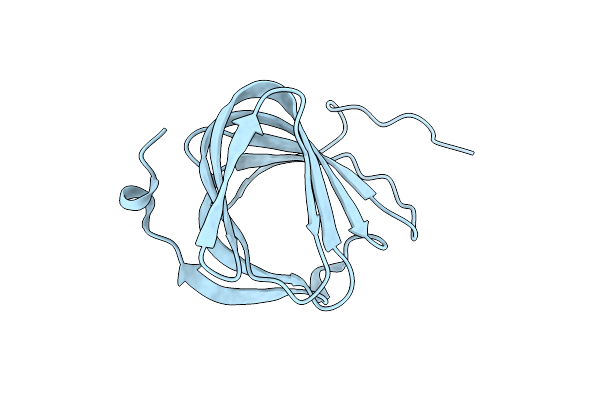
Method: X-RAY DIFFRACTION Resolution:1.60 Å Release Date: 2018-03-28 Classification: STRUCTURAL PROTEIN |
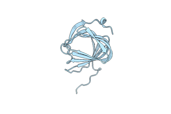 |
Roll Out The Beta-Barrel: Structure And Mechanism Of Pac13, A Unique Nucleoside Dehydratase
Organism: Streptomyces coeruleorubidus
Method: X-RAY DIFFRACTION Resolution:1.55 Å Release Date: 2018-03-28 Classification: STRUCTURAL PROTEIN |
 |
Organism: Streptomyces coeruleorubidus
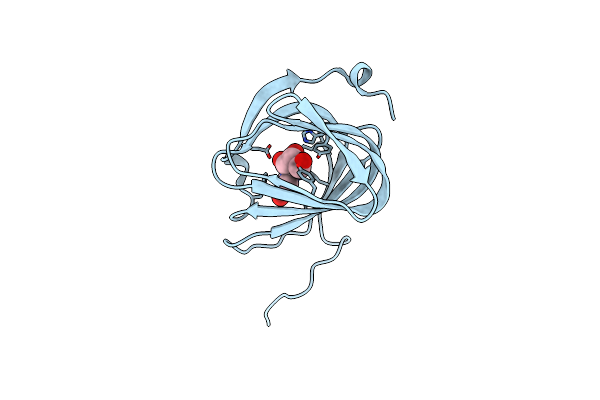
Method: X-RAY DIFFRACTION Resolution:1.60 Å Release Date: 2017-08-23 Classification: BIOSYNTHETIC PROTEIN Ligands: URI |
 |
Organism: Streptomyces coeruleorubidus
Method: X-RAY DIFFRACTION Resolution:1.78 Å Release Date: 2017-08-23 Classification: BIOSYNTHETIC PROTEIN Ligands: UUA |
 |
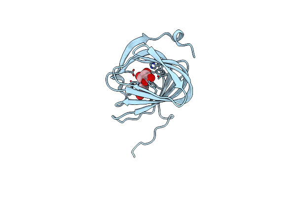
Organism: Streptomyces coeruleorubidus
Method: X-RAY DIFFRACTION Resolution:1.78 Å Release Date: 2017-08-23 Classification: BIOSYNTHETIC PROTEIN Ligands: EDO, URI |
 |
Organism: Streptomyces coeruleorubidus
Method: X-RAY DIFFRACTION Resolution:1.59 Å Release Date: 2017-08-23 Classification: BIOSYNTHETIC PROTEIN Ligands: URI |
 |
Organism: Streptomyces coeruleorubidus
Method: X-RAY DIFFRACTION Resolution:1.60 Å Release Date: 2017-08-23 Classification: BIOSYNTHETIC PROTEIN Ligands: URI |
 |
NA
Organism: Streptomyces coeruleorubidus
Method: Alphafold Release Date: Classification: NA Ligands: NA |