
Search Count: 23
All
Selected
 |
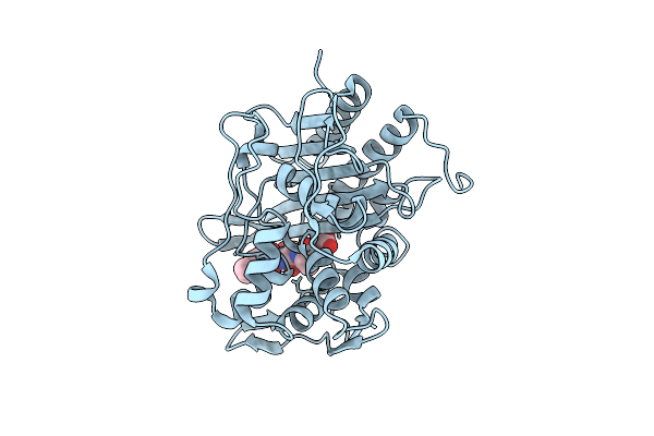
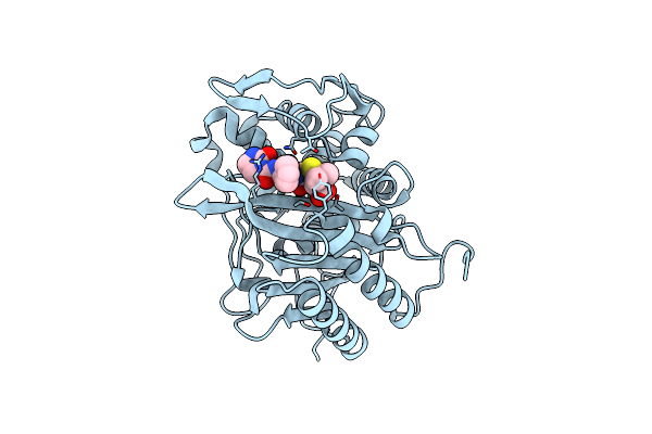
Crystal Structure Of The Transpeptidase Domain Of Pbp2 From The Neisseria Gonorrhoeae Cephalosporin-Resistant Strain H041 In Complex With Boronate Inhibitor Vnrx-14079
Organism: Neisseria gonorrhoeae
Method: X-RAY DIFFRACTION Resolution:2.10 Å Release Date: 2026-02-04 Classification: LIGASE Ligands: A1C02 |
 |
Organism: Homo sapiens
Method: ELECTRON MICROSCOPY Release Date: 2025-10-01 Classification: LIGASE Ligands: AMP, ZN |
 |
Organism: Homo sapiens
Method: ELECTRON MICROSCOPY Resolution:2.70 Å Release Date: 2025-10-01 Classification: LIGASE Ligands: ZN, AMP |
 |
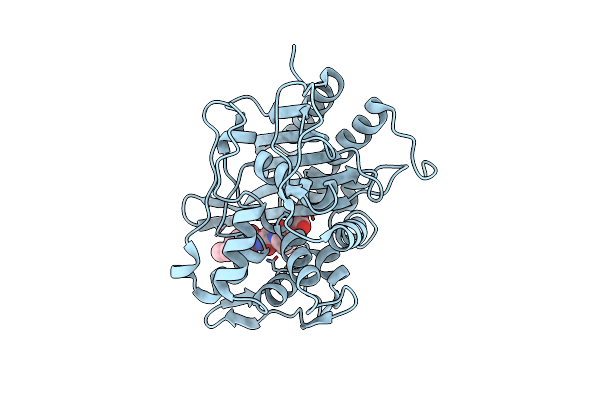
Crystal Structure Of The Transpeptidase Domain Of Pbp2 From The Neisseria Gonorrhoeae Cephalosporin Decreased Susceptibility Strain 35/02 In Complex With Boronate Inhibitor Vnrx-6884
Organism: Neisseria gonorrhoeae 35/02
Method: X-RAY DIFFRACTION Resolution:1.89 Å Release Date: 2025-01-22 Classification: LIGASE Ligands: A1BJC |
 |
Crystal Structure Of The Transpeptidase Domain Of Pbp2 From The Neisseria Gonorrhoeae Cephalosporin Decreased Susceptibility Strain 35/02 In Complex With Boronate Inhibitor Vnrx-6752
Organism: Neisseria gonorrhoeae 35/02
Method: X-RAY DIFFRACTION Resolution:2.61 Å Release Date: 2025-01-22 Classification: LIGASE Ligands: A1BJB |
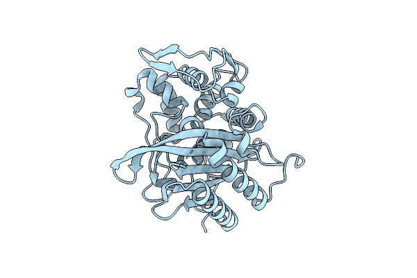 |
Crystal Structure Of The Transpeptidase Domain Of A S310A Mutant Of Pbp2 From Neisseria Gonorrhoeae Strain H041
Organism: Neisseria gonorrhoeae
Method: X-RAY DIFFRACTION Resolution:1.90 Å Release Date: 2024-03-20 Classification: HYDROLASE |
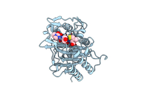 |
Crystal Structure Of Transpeptidase Domain Of Pbp2 From Neisseria Gonorrhoeae Cephalosporin-Resistant Strain H041 In Complex With Cefoperazone
Organism: Neisseria gonorrhoeae
Method: X-RAY DIFFRACTION Resolution:1.80 Å Release Date: 2024-03-20 Classification: HYDROLASE Ligands: A1ADP |
 |
Crystal Structure Of Transpeptidase Domain Of Pbp2 From Neisseria Gonorrhoeae Cephalosporin-Resistant Strain H041 Acylated By Piperacillin
Organism: Neisseria gonorrhoeae
Method: X-RAY DIFFRACTION Resolution:2.00 Å Release Date: 2024-03-20 Classification: HYDROLASE Ligands: JPP, PEG |
 |
Crystal Structure Of Transpeptidase Domain Of Pbp2 From Neisseria Gonorrhoeae Cephalosporin-Resistant Strain H041 In Complex With Azlocillin
Organism: Neisseria gonorrhoeae
Method: X-RAY DIFFRACTION Resolution:2.40 Å Release Date: 2024-03-20 Classification: HYDROLASE Ligands: 59H |
 |
Organism: Homo sapiens
Method: X-RAY DIFFRACTION Resolution:1.80 Å Release Date: 2022-08-03 Classification: Blood Clotting, Hydrolase Ligands: OQ6 |
 |
Organism: Homo sapiens
Method: X-RAY DIFFRACTION Resolution:1.80 Å Release Date: 2022-08-03 Classification: BLOOD CLOTTING, Hydrolase Ligands: OQI, CIT |
 |
Organism: Homo sapiens
Method: X-RAY DIFFRACTION Resolution:1.47 Å Release Date: 2022-08-03 Classification: BLOOD CLOTTING, hydrolase Ligands: OQO, CIT |
 |
Organism: Homo sapiens
Method: X-RAY DIFFRACTION Resolution:1.63 Å Release Date: 2022-08-03 Classification: BLOOD CLOTTING, Hydrolase Ligands: OR0, CIT |
 |
Organism: Homo sapiens
Method: X-RAY DIFFRACTION Resolution:1.59 Å Release Date: 2022-08-03 Classification: BLOOD CLOTTING, Hydrolase Ligands: CIT, ORF |
 |
Organism: Homo sapiens
Method: X-RAY DIFFRACTION Resolution:1.70 Å Release Date: 2022-08-03 Classification: BLOOD CLOTTING, Hydrolase Ligands: CIT, ORU |
 |
Organism: Homo sapiens
Method: X-RAY DIFFRACTION Resolution:1.68 Å Release Date: 2022-08-03 Classification: BLOOD CLOTTING, Hydrolase Ligands: CIT, OSX |
 |
Organism: Homo sapiens
Method: X-RAY DIFFRACTION Resolution:1.51 Å Release Date: 2022-08-03 Classification: BLOOD CLOTTING, Hydrolase Ligands: CIT, OT7 |
 |
Organism: Homo sapiens
Method: X-RAY DIFFRACTION Resolution:1.52 Å Release Date: 2022-08-03 Classification: BLOOD CLOTTING, Hydrolase Ligands: CIT, OTL |
 |
Organism: Homo sapiens
Method: X-RAY DIFFRACTION Resolution:1.73 Å Release Date: 2022-08-03 Classification: BLOOD CLOTTING, Hydrolase Ligands: CIT, OTX |
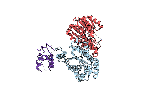 |
Structure Of Acyl Carrier Protein Bound To Fabi, The Enoyl Reductase From Escherichia Coli
Organism: Escherichia coli
Method: X-RAY DIFFRACTION Resolution:2.70 Å Release Date: 2006-10-17 Classification: OXIDOREDUCTASE/BIOSYNTHETIC PROTEIN |