
Search Count: 28
 |
Organism: Homo sapiens
Method: X-RAY DIFFRACTION Resolution:1.79 Å Release Date: 2024-07-03 Classification: APOPTOSIS Ligands: A1H2C |
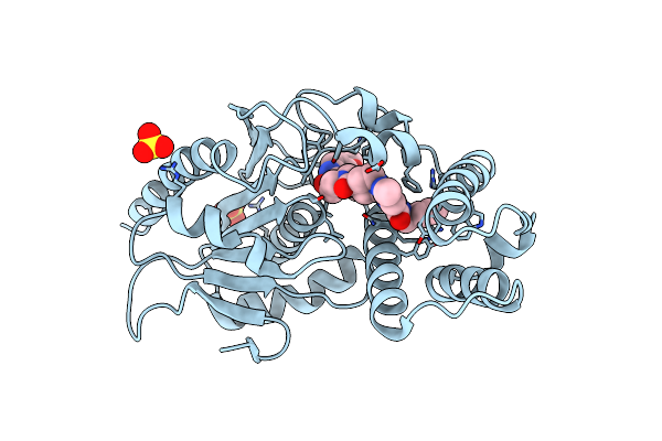 |
Crystal Structure Of Mtb Pks13 Thioesterase Domain In Complex With Inhibitor X20419
Organism: Mycobacterium tuberculosis
Method: X-RAY DIFFRACTION Resolution:2.20 Å Release Date: 2024-04-24 Classification: BIOSYNTHETIC PROTEIN Ligands: JR0, SO4 |
 |
Crystal Structure Of Mtb Pks13 Thioesterase Domain In Complex With Inhibitor X20403
Organism: Mycobacterium tuberculosis h37rv
Method: X-RAY DIFFRACTION Resolution:2.00 Å Release Date: 2024-04-24 Classification: BIOSYNTHETIC PROTEIN Ligands: JS9, SO4 |
 |
Crystal Structure Of Mtb Pks13 Thioesterase Domain In Complex With Inhibitor X20404
Organism: Mycobacterium tuberculosis
Method: X-RAY DIFFRACTION Resolution:2.10 Å Release Date: 2024-04-24 Classification: BIOSYNTHETIC PROTEIN Ligands: K6C, SO4 |
 |
Crystal Structure Of Mtb Pks13 Thioesterase Domain In Complex With Inhibitor X20348
Organism: Mycobacterium tuberculosis
Method: X-RAY DIFFRACTION Resolution:2.35 Å Release Date: 2024-04-24 Classification: BIOSYNTHETIC PROTEIN Ligands: QU0, SO4 |
 |
Organism: Homo sapiens
Method: ELECTRON MICROSCOPY Release Date: 2023-11-29 Classification: MEMBRANE PROTEIN Ligands: ACP, MG, NAG |
 |
Organism: Homo sapiens
Method: ELECTRON MICROSCOPY Release Date: 2023-11-29 Classification: MEMBRANE PROTEIN Ligands: ADP, ALF, MG, NAG |
 |
Organism: Homo sapiens
Method: ELECTRON MICROSCOPY Release Date: 2023-11-29 Classification: MEMBRANE PROTEIN Ligands: MG, ALF, NAG |
 |
Cryo-Em Structure Of Atp8B1-Cdc50A In E2P Autoinhibited "Closed" Conformation
Organism: Homo sapiens
Method: ELECTRON MICROSCOPY Release Date: 2023-11-29 Classification: MEMBRANE PROTEIN Ligands: NAG, MG, IP9 |
 |
Cryo-Em Structure Of Atp8B1-Cdc50A In E2P Autoinhibited "Open" Conformation
Organism: Homo sapiens
Method: ELECTRON MICROSCOPY Release Date: 2023-11-29 Classification: MEMBRANE PROTEIN Ligands: MG, IP9, NAG |
 |
Cryo-Em Structure Of Atp8B1-Cdc50A In E2P Active Conformation With Bound Pc
Organism: Homo sapiens
Method: ELECTRON MICROSCOPY Release Date: 2023-11-29 Classification: MEMBRANE PROTEIN Ligands: BEF, MG, POV, IP9, NAG |
 |
Organism: Homo sapiens
Method: ELECTRON MICROSCOPY Release Date: 2023-11-29 Classification: MEMBRANE PROTEIN Ligands: VN4, MG, D39, NAG |
 |
Organism: Homo sapiens
Method: ELECTRON MICROSCOPY Release Date: 2023-11-29 Classification: MEMBRANE PROTEIN Ligands: VN4, MG, POV, NAG |
 |
Organism: Homo sapiens
Method: ELECTRON MICROSCOPY Release Date: 2023-11-29 Classification: MEMBRANE PROTEIN Ligands: VN4, MG, PIE, NAG |
 |
Cryo-Em Map Of The Wt Kdpfabc Complex In The E1-P Tight Conformation, Stabilised With The Inhibitor Orthovanadate
Organism: Escherichia coli
Method: ELECTRON MICROSCOPY Release Date: 2022-11-16 Classification: MEMBRANE PROTEIN Ligands: K, CDL, VO4 |
 |
Cryo-Em Map Of The Wt Kdpfabc Complex In The E1-P Tight Conformation, Under Turnover Conditions
Organism: Escherichia coli
Method: ELECTRON MICROSCOPY Release Date: 2022-11-16 Classification: MEMBRANE PROTEIN Ligands: K, CDL |
 |
Cryo-Em Map Of The Wt Kdpfabc Complex In The E1_Atpearly Conformation, Under Turnover Conditions
Organism: Escherichia coli
Method: ELECTRON MICROSCOPY Release Date: 2022-11-16 Classification: MEMBRANE PROTEIN Ligands: K, CDL, ATP |
 |
Cryo-Em Structure Of The Kdpfabc Complex In A Nucleotide-Free E1 Conformation Loaded With K+
Organism: Escherichia coli
Method: ELECTRON MICROSCOPY Release Date: 2022-11-16 Classification: MEMBRANE PROTEIN Ligands: K, CDL |
 |
Cryo-Em Structure Of The Kdpfabc Complex In A Nucleotide-Free E1 Conformation Loaded With K+
Organism: Escherichia coli
Method: ELECTRON MICROSCOPY Release Date: 2022-11-16 Classification: MEMBRANE PROTEIN Ligands: K, CDL |
 |
Cryo-Em Structure Of The Kdpfabc Complex In A Nucleotide-Free E1 Conformation Loaded With K+
Organism: Escherichia coli
Method: ELECTRON MICROSCOPY Release Date: 2022-11-16 Classification: MEMBRANE PROTEIN Ligands: K, CDL |