
Search Count: 28
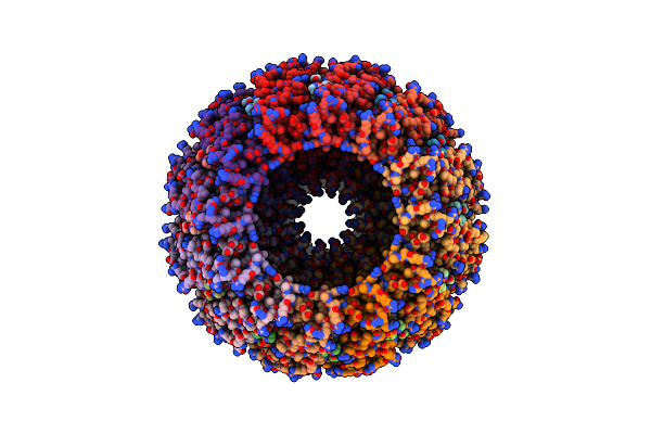 |
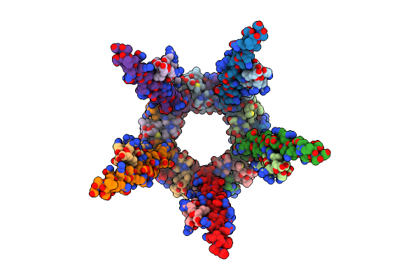
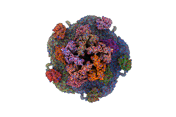
Herpes Simplex Virus Type 1 (Hsv-1) A-Capsid Pul6 Portal Protein, Dodecameric Complex
Organism: Human alphaherpesvirus 1 strain kos
Method: ELECTRON MICROSCOPY Release Date: 2025-06-11 Classification: VIRAL PROTEIN |
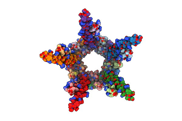 |
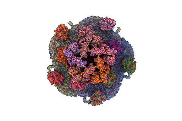
Organism: Human alphaherpesvirus 1 strain kos
Method: ELECTRON MICROSCOPY Release Date: 2025-06-11 Classification: VIRAL PROTEIN |
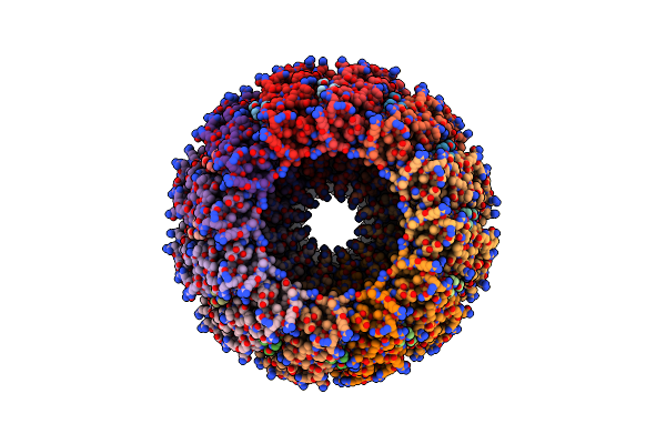 |
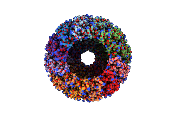
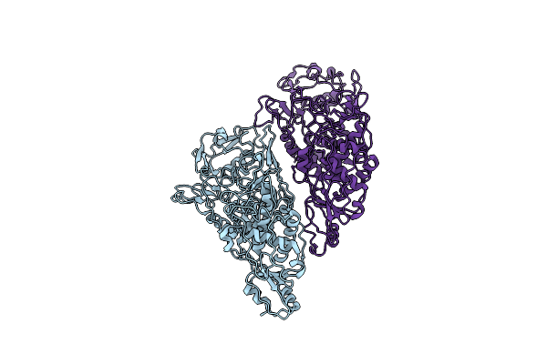
Herpes Simplex Virus Type 1 (Hsv-1) B-Capsid Pul6 Portal Protein, Dodecameric Complex
Organism: Human alphaherpesvirus 1 strain kos
Method: ELECTRON MICROSCOPY Release Date: 2025-05-28 Classification: VIRAL PROTEIN |
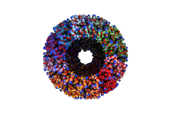 |
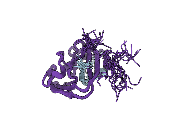
Herpes Simplex Virus Type 1 (Hsv-1) D-Capsid Pul6 Portal Protein, Dodecameric Complex
Organism: Human alphaherpesvirus 1 strain kos
Method: ELECTRON MICROSCOPY Release Date: 2025-05-28 Classification: VIRAL PROTEIN |
 |
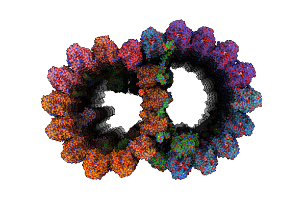
Herpes Simplex Virus Type 1 (Hsv-1) D-Capsid Pul6 Portal Protein Turrets, Decamer
Organism: Human alphaherpesvirus 1 strain kos
Method: ELECTRON MICROSCOPY Release Date: 2025-05-28 Classification: VIRAL PROTEIN |
 |
Herpes Simplex Virus Type 1 (Hsv-1) C-Capsid Pul6 Portal Protein, Dodecameric Complex
Organism: Human alphaherpesvirus 1 strain kos
Method: ELECTRON MICROSCOPY Release Date: 2025-05-28 Classification: VIRAL PROTEIN |
 |
Organism: Trichomonas vaginalis g3
Method: ELECTRON MICROSCOPY Release Date: 2025-05-14 Classification: STRUCTURAL PROTEIN Ligands: GDP, GTP, MG |
 |
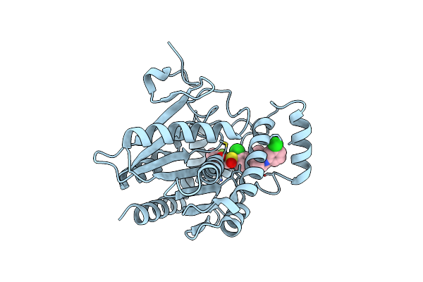
Complex Crystal Structure Of Mutant Human Monoglyceride Lipase With Compound 5D
Organism: Homo sapiens
Method: X-RAY DIFFRACTION Resolution:1.51 Å Release Date: 2024-01-31 Classification: HYDROLASE Ligands: F28, EDO |
 |
Complex Crystal Structure Of Mutant Human Monoglyceride Lipase With Compound 5L
Organism: Homo sapiens
Method: X-RAY DIFFRACTION Resolution:1.55 Å Release Date: 2024-01-31 Classification: HYDROLASE Ligands: EJI, EDO |
 |
Complex Crystal Structure Of Mutant Human Monoglyceride Lipase With Compound 5R
Organism: Homo sapiens
Method: X-RAY DIFFRACTION Resolution:1.73 Å Release Date: 2024-01-31 Classification: HYDROLASE Ligands: EFH, EDO |
 |
Organism: Homo sapiens
Method: X-RAY DIFFRACTION Resolution:1.55 Å Release Date: 2023-08-23 Classification: HYDROLASE Ligands: NUX |
 |
Organism: Golden shiner reovirus
Method: ELECTRON MICROSCOPY Release Date: 2023-03-22 Classification: VIRUS Ligands: ZN |
 |
Organism: Golden shiner reovirus
Method: ELECTRON MICROSCOPY Release Date: 2023-03-08 Classification: VIRUS Ligands: ZN |
 |
Organism: Trichomonas vaginalis virus 2
Method: ELECTRON MICROSCOPY Release Date: 2021-04-07 Classification: VIRAL PROTEIN |
 |
Organism: Trichomonas vaginalis virus 2
Method: ELECTRON MICROSCOPY Release Date: 2021-04-07 Classification: VIRAL PROTEIN |
 |
Organism: Enterobacteria phage t7
Method: SOLUTION NMR Release Date: 2018-09-19 Classification: SPLICING |
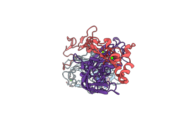 |
Crystal Structure Of A Ternary Complex Of Factor Viia/Tissue Factor/Pyrazinone Inhibitor
Organism: Homo sapiens
Method: X-RAY DIFFRACTION Resolution:2.00 Å Release Date: 2005-05-03 Classification: HYDROLASE Ligands: CA, MG, PY3 |
 |
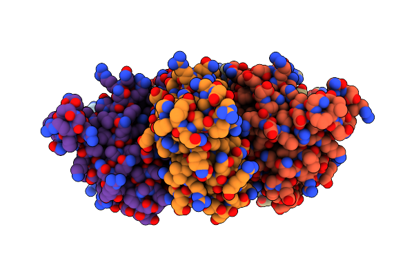
Murine Inducible Nitric Oxide Synthase Oxygenase Dimer, Tetrahydrobiopterin And 4R-Fluoro-N6-Ethanimidoyl-L-Lysine
Organism: Mus musculus
Method: X-RAY DIFFRACTION Resolution:2.30 Å Release Date: 2004-10-05 Classification: OXIDOREDUCTASE Ligands: SO4, HEM, H4B, I58 |
 |
Structure Of A Hexameric N-Terminal Domain From Murine Leukemia Virus Capsid
Organism: Akr (endogenous) murine leukemia virus
Method: X-RAY DIFFRACTION Resolution:1.85 Å Release Date: 2004-10-05 Classification: VIRAL PROTEIN |
 |
Organism: Homo sapiens
Method: SOLUTION NMR Release Date: 2001-12-12 Classification: HYDROLASE/HYDROLASE INHIBITOR Ligands: ZN, CA, I52 |