
Search Count: 12
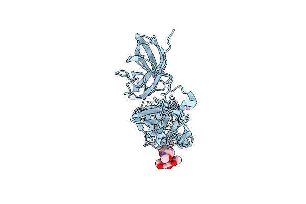 |
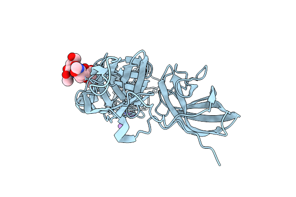
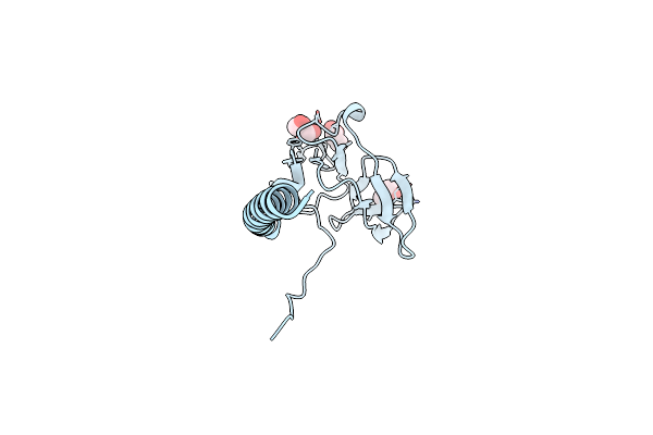
Structure Of Hantavirus Envelope Glycoprotein Gc In Postfusion Conformation In Presence Of 600 Mm Kcl
Organism: Hantaan virus
Method: X-RAY DIFFRACTION Resolution:1.40 Å Release Date: 2016-09-14 Classification: VIRAL PROTEIN Ligands: K, NA |
 |
Organism: Hantaan virus (strain 76-118), Homo sapiens
Method: X-RAY DIFFRACTION Resolution:3.00 Å Release Date: 2016-09-14 Classification: VIRAL PROTEIN Ligands: NAG, NCO |
 |
Structure Of Hantavirus Envelope Glycoprotein Gc In Postfusion Conformation
Organism: Hantaan virus
Method: X-RAY DIFFRACTION Resolution:1.60 Å Release Date: 2016-09-14 Classification: VIRAL PROTEIN Ligands: MES, NA |
 |
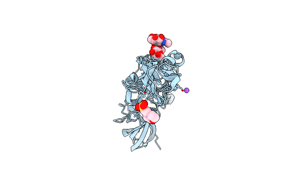
Structure Of Hantavirus Envelope Glycoprotein Gc In Postfusion Conformation In Presence Of 100 Mm Kcl
Organism: Hantaan virus
Method: X-RAY DIFFRACTION Resolution:1.80 Å Release Date: 2016-09-14 Classification: VIRAL PROTEIN |
 |
Structure Of Hantavirus Envelope Glycoprotein Gc In Postfusion Conformation In Presence Of 200 Mm Kcl
Organism: Hantaan virus
Method: X-RAY DIFFRACTION Resolution:1.70 Å Release Date: 2016-09-14 Classification: VIRAL PROTEIN Ligands: K, NA |
 |
Structure Of Hantavirus Envelope Glycoprotein Gc In Postfusion Conformation In Presence Of 300 Mm Kcl
Organism: Hantaan virus
Method: X-RAY DIFFRACTION Resolution:1.60 Å Release Date: 2016-09-14 Classification: VIRAL PROTEIN Ligands: K, NA |
 |
Structure Of Hantavirus Envelope Glycoprotein Gc In Postfusion Conformation In Presence Of 500 Mm Kcl
Organism: Hantaan virus
Method: X-RAY DIFFRACTION Resolution:1.50 Å Release Date: 2016-09-14 Classification: VIRAL PROTEIN Ligands: K, NA |
 |
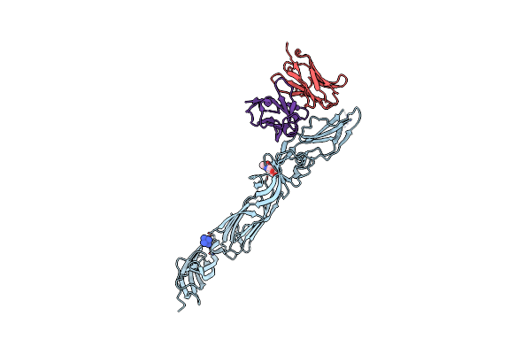
Crystal Structure Of The T1L Reovirus Attachment Protein Sigma1 In Complex With Junctional Adhesion Molecule-A
Organism: Mammalian orthoreovirus 1, Homo sapiens
Method: X-RAY DIFFRACTION Resolution:3.20 Å Release Date: 2015-04-01 Classification: VIRAL PROTEIN |
 |
Organism: Mammalian orthoreovirus 1 lang
Method: X-RAY DIFFRACTION Resolution:2.20 Å Release Date: 2015-04-01 Classification: VIRAL PROTEIN Ligands: CL, MG, ACT, GOL |
 |
Organism: Enterobacteria phage p22
Method: X-RAY DIFFRACTION Resolution:1.65 Å Release Date: 2011-05-04 Classification: HYDROLASE Ligands: GOL, CA, PE4, SO4 |
 |
Headbinding Domain Of Phage P22 Tailspike C-Terminally Fused To Isoleucine Zipper Piigcn4 (Chimera I)
Organism: Enterobacteria phage p22, Saccharomyces cerevisiae
Method: X-RAY DIFFRACTION Resolution:2.05 Å Release Date: 2009-02-10 Classification: VIRAL PROTEIN |
 |
Mutant Y108Wdel Of The Headbinding Domain Of Phage P22 Tailspike C- Terminally Fused To Isoleucine Zipper Piigcn4 (Chimera Ii)
Organism: Enterobacteria phage p22, Saccharomyces cerevisiae
Method: X-RAY DIFFRACTION Resolution:1.80 Å Release Date: 2009-02-10 Classification: VIRAL PROTEIN Ligands: GOL |