
Search Count: 10
 |
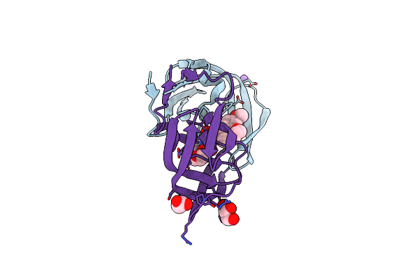
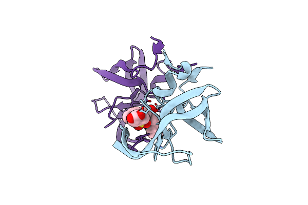
A Hydroxymethyl Functionality At The 4-Position Of The 2-Phenyloxazole Moiety Of Hiv-1 Protease Inhibitors Involving The P2' Ligands
Organism: Human immunodeficiency virus 1
Method: X-RAY DIFFRACTION Resolution:1.30 Å Release Date: 2017-11-22 Classification: HYDROLASE/HYDROLASE INHIBITOR Ligands: G53, NA, CL |
 |
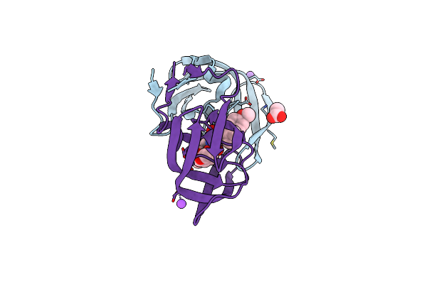
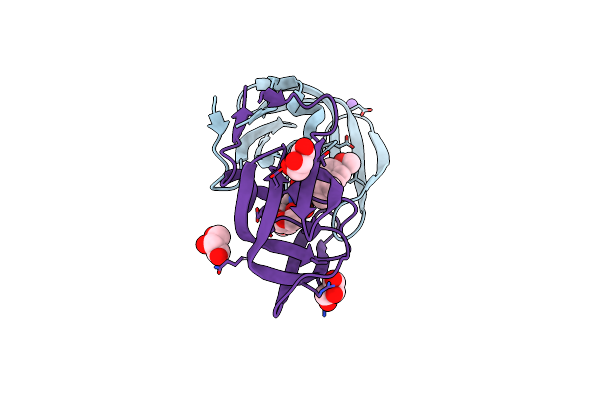
A Novel 13-Ring Macrocyclic Hiv-1 Protease Inhibitors Involving The P1'-P2' Ligands
Organism: Human immunodeficiency virus 1
Method: X-RAY DIFFRACTION Resolution:1.27 Å Release Date: 2017-10-11 Classification: hydrolase/hydrolase inhibitor |
 |
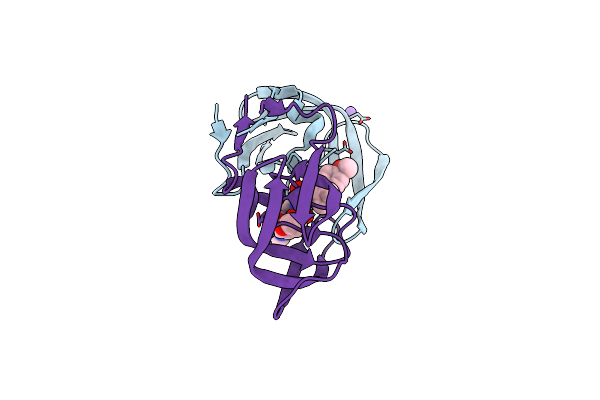
Crystal Structure Of Hiv-1 Protease Inhibitors Containing Substituted Fused-Tetrahydropyranyl Tetrahydrofuran As P2-Ligand Grl-004-11A
Organism: Human immunodeficiency virus 1
Method: X-RAY DIFFRACTION Resolution:1.22 Å Release Date: 2015-10-28 Classification: HYDROLASE Ligands: 5B7, NA, CL, ACT |
 |
Crystal Structure Of Hiv-1 Protease Inhibitor Grl-105-11A Containing Substituted Fused-Tetrahydropyranyl Tetrahydrofuran As P2-Ligand
Organism: Human immunodeficiency virus 1
Method: X-RAY DIFFRACTION Resolution:1.62 Å Release Date: 2015-10-28 Classification: HYDROLASE Ligands: 5B5, NA, CL |
 |
Organism: Human immunodeficiency virus type 1 (bru isolate)
Method: X-RAY DIFFRACTION Resolution:1.95 Å Release Date: 2013-07-24 Classification: HYDROLASE/HYDROLASE INHIBITOR Ligands: 017 |
 |
Crystal Structure Of Wild Type Hiv-1 Protease In Complex With Non-Peptidic Inhibitor, Grl007
Organism: Human immunodeficiency virus type 1
Method: X-RAY DIFFRACTION Resolution:1.96 Å Release Date: 2013-07-24 Classification: HYDROLASE/HYDROLASE INHIBITOR Ligands: G07 |
 |
Crystal Structure Of Wild Type Hiv-1 Protease In Complex With Non-Peptidic Inhibitor, Grl008
Organism: Human immunodeficiency virus type 1
Method: X-RAY DIFFRACTION Resolution:1.75 Å Release Date: 2013-07-24 Classification: HYDROLASE/HYDROLASE INHIBITOR Ligands: G08 |
 |
Crystal Structure Of Wild-Type Hiv-1 Protease With Cyclopentyltetrahydro- Furanyl Urethanes As P2-Ligand, Grl-0249A
Organism: Human immunodeficiency virus type 1
Method: X-RAY DIFFRACTION Resolution:1.23 Å Release Date: 2012-03-21 Classification: hydrolase/hydrolase inhibitor |
 |
Hiv-1 Wild Type Protease With A Substituted Bis-Tetrahydrofuran Inhibitor, Grl-044-10A
Organism: Human immunodeficiency virus 1
Method: X-RAY DIFFRACTION Resolution:1.40 Å Release Date: 2011-12-21 Classification: HYDROLASE/HYDROLASE INHIBITOR Ligands: G04, NA, CL, GOL |
 |
Crystal Structure Of Wild-Type Hiv-1 Protease With C3-Substituted Hexahydrocyclopentafuranyl Urethane As P2-Ligand, Grl-0489A
Organism: Human immunodeficiency virus type 1
Method: X-RAY DIFFRACTION Resolution:1.45 Å Release Date: 2011-08-17 Classification: HYDROLASE/HYDROLASE INHIBITOR Ligands: CL, G89 |