
Search Count: 117
 |
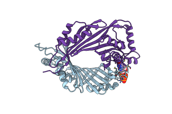
Crystal Structure Of De Novo Designed Metal-Controlled Heterodimer Of Mutant B1 Immunoglobulin-Binding Domain Of Streptococcal Protein G Mchet_A + Mchet_B
Organism: Streptococcus pyogenes
Method: X-RAY DIFFRACTION Resolution:1.43 Å Release Date: 2024-10-09 Classification: DE NOVO PROTEIN Ligands: ZN, CL |
 |
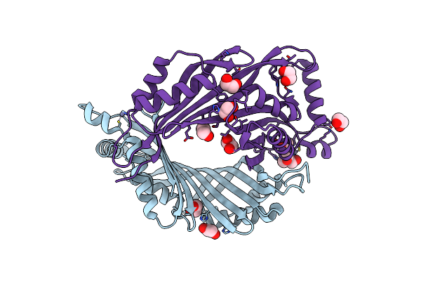
Crystal Structure Of De Novo Designed Metal-Controlled Heterodimer Of Mutant B1 Immunoglobulin-Binding Domain Of Streptococcal Protein G Mchet_A + Mchet_C
Organism: Streptococcus pyogenes
Method: X-RAY DIFFRACTION Resolution:1.36 Å Release Date: 2024-10-09 Classification: DE NOVO PROTEIN Ligands: ZN, CL, EDO |
 |
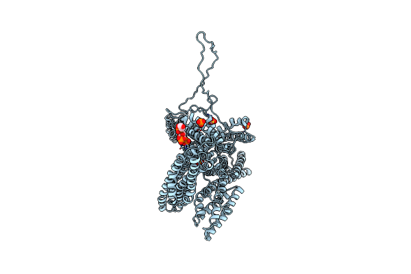
Organism: Drosophila melanogaster
Method: X-RAY DIFFRACTION Resolution:2.50 Å Release Date: 2024-02-07 Classification: MOTOR PROTEIN Ligands: MG, ADP |
 |
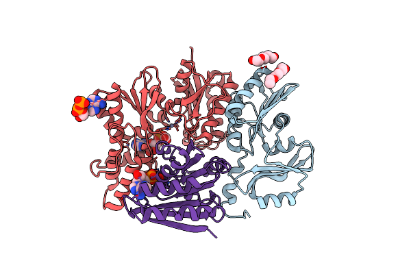
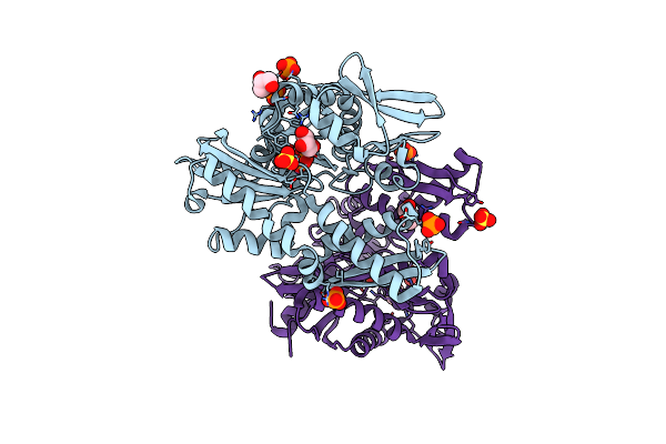
Organism: Thermotoga maritima msb8
Method: X-RAY DIFFRACTION Resolution:3.14 Å Release Date: 2023-04-26 Classification: RNA BINDING PROTEIN Ligands: TXA, ZN, MG, APC |
 |
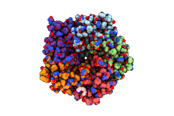
Organism: Saccharomyces cerevisiae s288c, Saccharomyces cerevisiae s288c
Method: X-RAY DIFFRACTION Resolution:2.94 Å Release Date: 2023-04-26 Classification: RNA BINDING PROTEIN Ligands: ZN, AMP, PGE, ATP, SO4, ACT |
 |
Organism: Gallus gallus
Method: X-RAY DIFFRACTION Resolution:3.00 Å Release Date: 2020-01-29 Classification: CELL ADHESION Ligands: SO4, KYG, PO4 |
 |
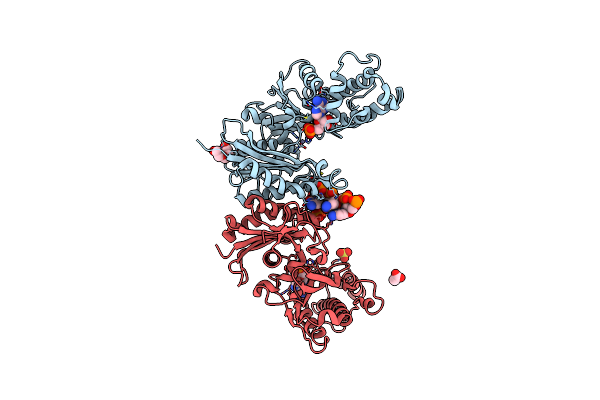
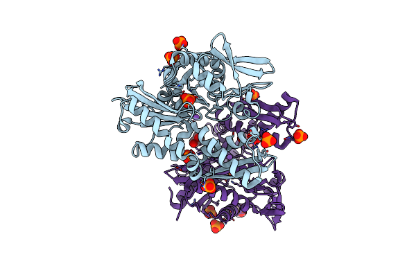
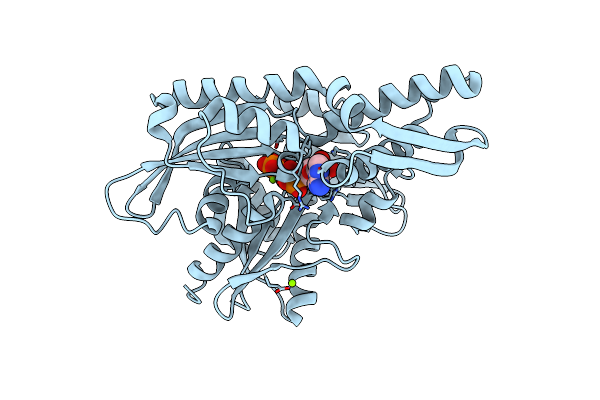
Crystal Structure Of Thermotoga Maritima Threonylcarbamoyladenosine Biosynthesis Complex Tsab2D2E2 Bound To Atp And Carboxy-Amp
Organism: Thermotoga maritima
Method: X-RAY DIFFRACTION Resolution:2.50 Å Release Date: 2019-05-22 Classification: BIOSYNTHETIC PROTEIN Ligands: PGE, AE3, KG4, ZN, ADP, ATP, MG |
 |
Crystal Structure Of Mutant B1 Immunoglobulin-Binding Domain Of Streptococcal Protein G (T16F, T18A, V21E, T25L, K28Y, V29I, K31R, Q32H, Y33L, N35K, D36H, N37Q)
Organism: Streptococcus
Method: X-RAY DIFFRACTION Resolution:1.40 Å Release Date: 2019-01-23 Classification: DE NOVO PROTEIN Ligands: ZN, CL |
 |
Crystal Structure Of B1 Immunoglobulin-Binding Domain Of Streptococcal Protein G (T16F, T18A, V21H, T25H, K28Y, V29I, K31R, Q32A, Y33L, N35K, D36A, N37Q)
Organism: Streptococcus
Method: X-RAY DIFFRACTION Resolution:1.40 Å Release Date: 2019-01-23 Classification: METAL BINDING PROTEIN Ligands: ZN, ACT, NA, CL, PO4, DPO |
 |
Crystal Structure Of De Novo Designed Metal-Controlled Dimer Of Mutant B1 Immunoglobulin-Binding Domain Of Streptococcal Protein G (L12H, T16L, V29H, Y33H, N37L)-Zinc
Organism: Streptococcus
Method: X-RAY DIFFRACTION Resolution:1.50 Å Release Date: 2019-01-23 Classification: METAL BINDING PROTEIN Ligands: ZN, CL |
 |
Crystal Structure Of De Novo Designed Metal-Controlled Dimer Of Mutant B1 Immunoglobulin-Binding Domain Of Streptococcal Protein G (L12H, T16L, V29H, Y33H, N37L)-Apo
Organism: Streptococcus
Method: X-RAY DIFFRACTION Resolution:1.70 Å Release Date: 2019-01-23 Classification: DE NOVO PROTEIN Ligands: MG, NA |
 |
Crystal Structure Of De Novo Designed Metal-Controlled Dimer Of B1 Immunoglobulin-Binding Domain Of Streptococcal Protein G (L12H, E15V, T16L, T18I, V29H, Y33H, N37L)-Zinc
Organism: Streptococcus
Method: X-RAY DIFFRACTION Resolution:1.34 Å Release Date: 2019-01-23 Classification: DE NOVO PROTEIN Ligands: ZN, CL, GOL, NA |
 |
Crystal Structure Of De Novo Designed Metal-Controlled Dimer Of Mutant B1 Immunoglobulin-Binding Domain Of Streptococcal Protein G (L12H, E15V, T16L, T18I, V29H, Y33H, N37L)-Apo
Organism: Streptococcus
Method: X-RAY DIFFRACTION Resolution:2.30 Å Release Date: 2019-01-23 Classification: METAL BINDING PROTEIN Ligands: MG |
 |
Mutant E97Q Crystal Structure Of Bacillus Subtilis Quef With A Disulfide Cys 55-99
Organism: Bacillus subtilis
Method: X-RAY DIFFRACTION Resolution:2.50 Å Release Date: 2017-03-29 Classification: OXIDOREDUCTASE Ligands: MG, PGE |
 |
Organism: Neisseria gonorrhoeae (strain atcc 700825 / fa 1090)
Method: X-RAY DIFFRACTION Resolution:2.77 Å Release Date: 2016-09-07 Classification: HYDROLASE, BIOSYNTHETIC PROTEIN Ligands: 8GT, ZN |
 |
Organism: Neisseria gonorrhoeae (strain atcc 700825 / fa 1090)
Method: X-RAY DIFFRACTION Resolution:1.90 Å Release Date: 2016-09-07 Classification: HYDROLASE, BIOSYNTHETIC PROTEIN Ligands: ZN, EDO, TRS, FMT |
 |
Organism: Homo sapiens
Method: X-RAY DIFFRACTION Resolution:1.86 Å Release Date: 2014-03-19 Classification: TRANSCRIPTION Ligands: PO4, CL, NA, GOL |
 |
Organism: Homo sapiens
Method: X-RAY DIFFRACTION Resolution:1.64 Å Release Date: 2014-03-19 Classification: TRANSCRIPTION |
 |
Organism: Homo sapiens
Method: X-RAY DIFFRACTION Resolution:1.90 Å Release Date: 2014-03-19 Classification: TRANSCRIPTION Ligands: ADP, MG, PO4 |
 |
Organism: Homo sapiens
Method: X-RAY DIFFRACTION Resolution:1.75 Å Release Date: 2014-03-19 Classification: TRANSCRIPTION Ligands: SO4, MG, CL, GOL, ACT |