
Search Count: 16
All
Selected
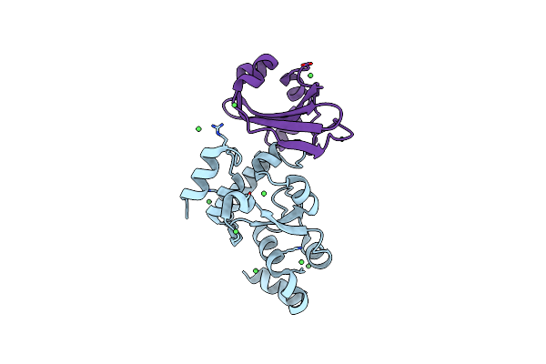 |
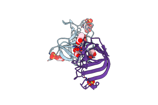
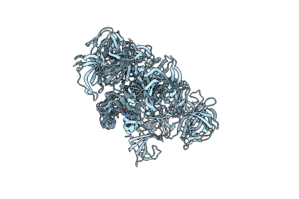
Organism: Staphylococcus aureus, Staphylococcus simulans
Method: X-RAY DIFFRACTION Resolution:2.10 Å Release Date: 2022-04-20 Classification: VIRAL PROTEIN Ligands: NI |
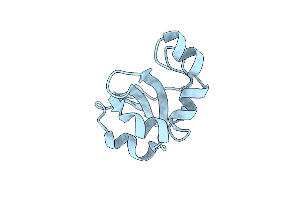 |
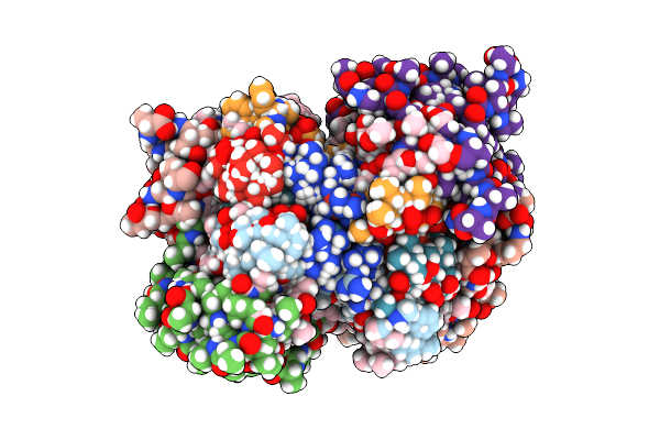
Organism: Staphylococcus simulans
Method: X-RAY DIFFRACTION Resolution:1.70 Å Release Date: 2022-04-20 Classification: VIRAL PROTEIN |
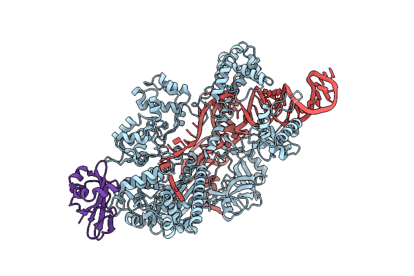 |
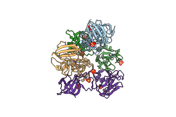
Organism: Staphylococcus aureus, Staphylococcus simulans
Method: X-RAY DIFFRACTION Resolution:4.21 Å Release Date: 2022-04-20 Classification: VIRAL PROTEIN/RNA |
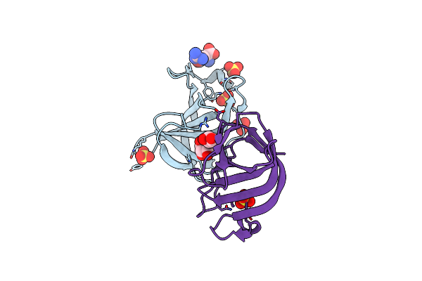 |
Organism: Staphylococcus simulans
Method: X-RAY DIFFRACTION Resolution:1.47 Å Release Date: 2021-02-17 Classification: CELL ADHESION Ligands: GOL, GGB, SO4 |
 |
Organism: Staphylococcus simulans
Method: X-RAY DIFFRACTION Resolution:0.84 Å Release Date: 2021-02-17 Classification: CELL ADHESION Ligands: BEZ, GOL, SO4, NA, MES |
 |
Organism: Staphylococcus simulans
Method: X-RAY DIFFRACTION Resolution:1.31 Å Release Date: 2021-02-17 Classification: CELL ADHESION Ligands: SAL, GOL, SO4 |
 |
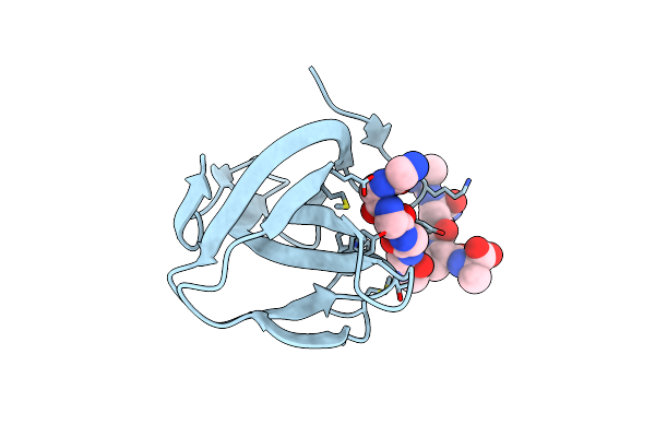
Solid-State Nmr Structure Of The D-Arg4,L10-Teixobactin - Lipid Ii Complex In Lipid Bilayers.
Organism: Eleftheria terrae, Staphylococcus simulans
Method: SOLID-STATE NMR Release Date: 2020-06-10 Classification: ANTIBIOTIC Ligands: 2PO, P1W |
 |
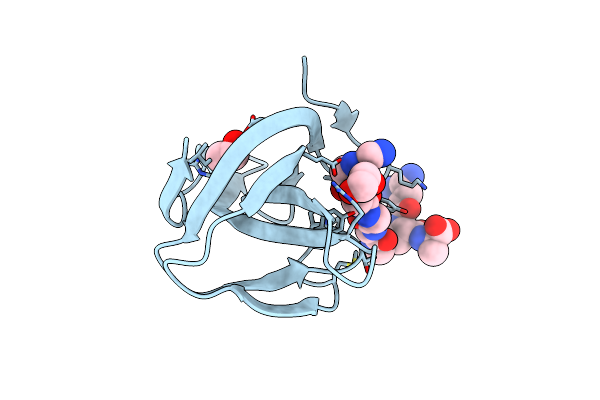
Organism: Staphylococcus simulans
Method: X-RAY DIFFRACTION Resolution:2.50 Å Release Date: 2019-10-16 Classification: PEPTIDE BINDING PROTEIN Ligands: K5T |
 |
Organism: Staphylococcus simulans
Method: X-RAY DIFFRACTION Resolution:1.43 Å Release Date: 2019-10-16 Classification: PEPTIDE BINDING PROTEIN Ligands: K5T, EDO |
 |
Organism: Staphylococcus simulans
Method: SOLUTION NMR Release Date: 2018-05-16 Classification: HYDROLASE Ligands: ZN |
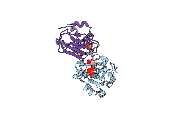 |
Catalytic Domain Of The Antimicrobial Peptidase Lysostaphin From Staphylococcus Simulans Crystallized In The Presence Of Phosphate
Organism: Staphylococcus simulans bv. staphylolyticus
Method: X-RAY DIFFRACTION Resolution:1.26 Å Release Date: 2014-07-16 Classification: HYDROLASE Ligands: ZN, PO4, GOL |
 |
Organism: Staphylococcus simulans
Method: X-RAY DIFFRACTION Resolution:3.50 Å Release Date: 2014-07-09 Classification: HYDROLASE Ligands: ZN, SO4 |
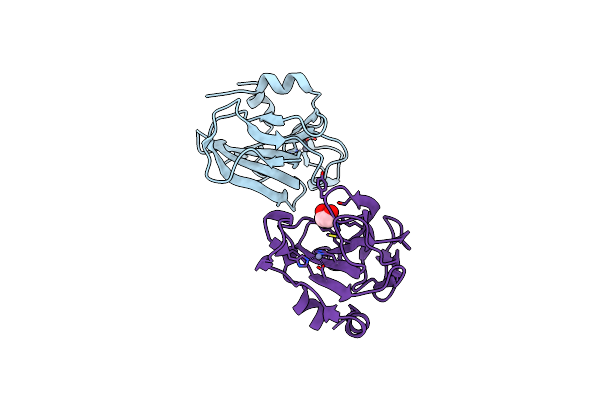 |
Catalytic Domain Of The Antimicrobial Peptidase Lysostaphin From Staphylococcus Simulans Crystallized In The Absence Of Phosphate
Organism: Staphylococcus simulans
Method: X-RAY DIFFRACTION Resolution:1.78 Å Release Date: 2014-07-09 Classification: HYDROLASE Ligands: ZN, EDO |
 |
NA
|
 |
NA
|
 |
NA
|