
Search Count: 16
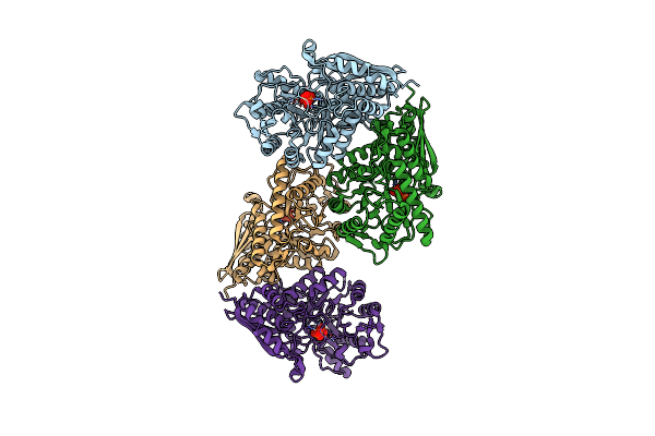 |
Organism: Streptococcus pyogenes mgas10394
Method: X-RAY DIFFRACTION Resolution:2.15 Å Release Date: 2014-02-05 Classification: LYASE Ligands: PO4 |
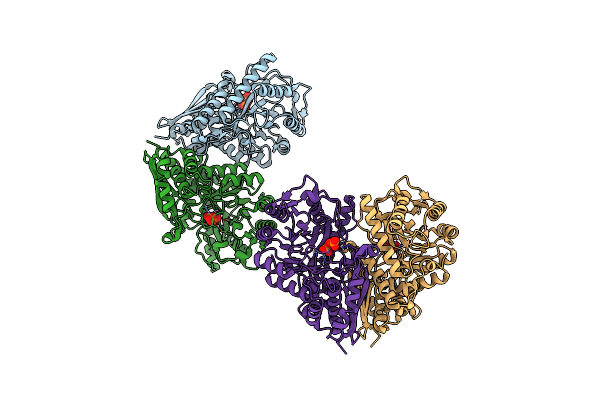 |
Organism: Streptococcus pyogenes mgas10394
Method: X-RAY DIFFRACTION Resolution:2.10 Å Release Date: 2014-02-05 Classification: LYASE Ligands: PO4 |
 |
Organism: Streptococcus pyogenes mgas10394
Method: X-RAY DIFFRACTION Resolution:2.90 Å Release Date: 2014-02-05 Classification: LYASE |
 |
Organism: Cryptococcus neoformans
Method: X-RAY DIFFRACTION Resolution:2.20 Å Release Date: 2012-10-24 Classification: OXIDOREDUCTASE Ligands: MOA, IMP, SO4 |
 |
Organism: Homo sapiens
Method: X-RAY DIFFRACTION Resolution:3.01 Å Release Date: 2011-09-14 Classification: IMMUNE SYSTEM Ligands: DTT |
 |
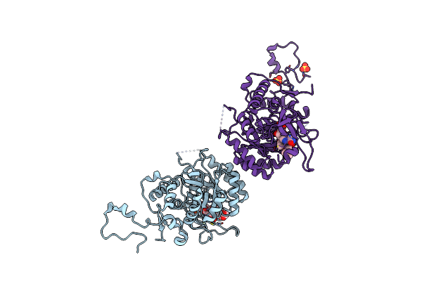
Structural Basis For High Arginine Specificity In Salmonella Typhimurium Periplasmic Binding Protein Stm4351.
Organism: Salmonella enterica subsp. enterica
Method: X-RAY DIFFRACTION Resolution:1.90 Å Release Date: 2011-05-11 Classification: ARGININE-BINDING PROTEIN Ligands: ARG, ZN, ACT, GOL |
 |
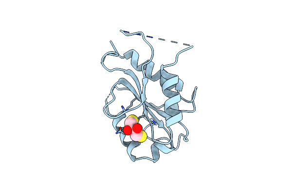
Organism: Salmonella enterica
Method: X-RAY DIFFRACTION Resolution:2.30 Å Release Date: 2010-07-28 Classification: HYDROLASE Ligands: ZN, PG4 |
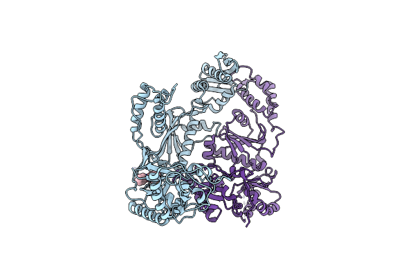 |
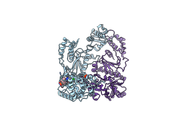
Organism: Hiv-1 m:b_hxb2r
Method: X-RAY DIFFRACTION Resolution:2.50 Å Release Date: 2008-08-12 Classification: TRANSFERASE Ligands: GFA |
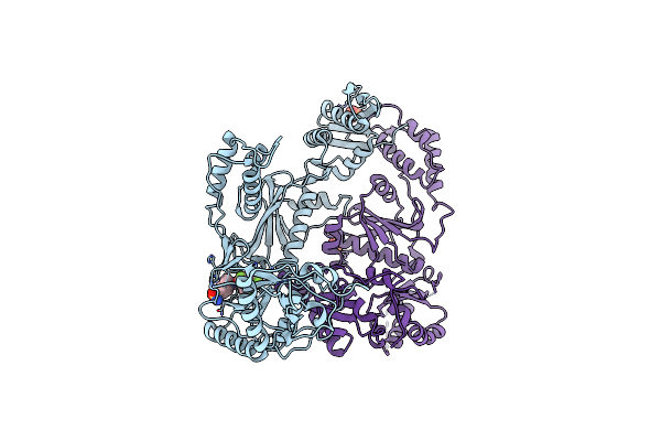 |
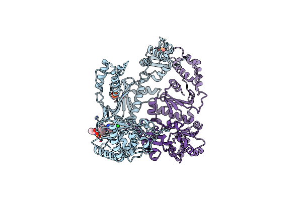
Organism: Hiv-1 m:b_hxb2r
Method: X-RAY DIFFRACTION Resolution:2.20 Å Release Date: 2008-08-12 Classification: TRANSFERASE Ligands: PO4, GWE |
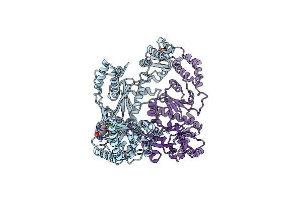 |
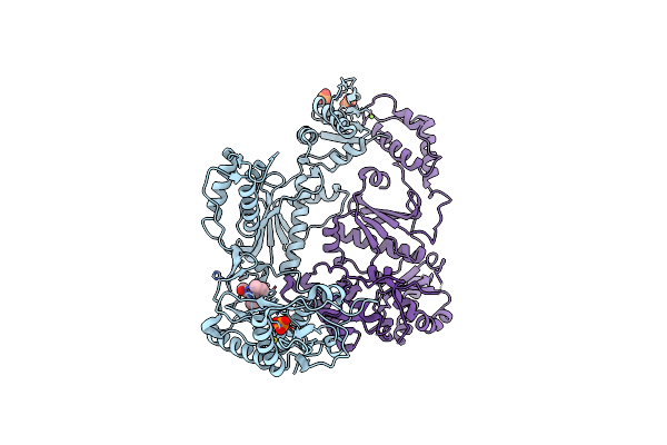
Crystal Structure Of Hiv-1 K103N Mutant Reverse Transcriptase In Complex With Gw564511.
Organism: Human immunodeficiency virus type 1
Method: X-RAY DIFFRACTION Resolution:3.10 Å Release Date: 2008-08-12 Classification: TRANSFERASE Ligands: PO4, GWE |
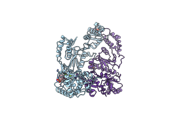 |
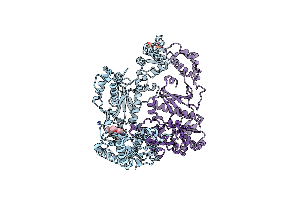
Crystal Structure Of Hiv-1 V106A And Y181C Mutant Reverse Transcriptase In Complex With Gw564511.
Organism: Human immunodeficiency virus type 1
Method: X-RAY DIFFRACTION Resolution:2.60 Å Release Date: 2008-08-12 Classification: TRANSFERASE Ligands: PO4, GWE |
 |
Crystal Structure Of K103N Mutant Hiv-1 Reverse Transcriptase In Complex With Gw678248.
Organism: Human immunodeficiency virus type 1
Method: X-RAY DIFFRACTION Resolution:2.90 Å Release Date: 2008-08-12 Classification: TRANSFERASE Ligands: PO4, GWJ |
 |
Crystal Structure Of L100I Mutant Hiv-1 Reverse Transcriptase In Complex With Gw695634.
Organism: Human immunodeficiency virus type 1
Method: X-RAY DIFFRACTION Resolution:2.50 Å Release Date: 2008-08-12 Classification: TRANSFERASE Ligands: PO4, GWI |
 |
Crystal Structure Of K101E Mutant Hiv-1 Reverse Transcriptase In Complex With Nevirapine
Organism: Human immunodeficiency virus 1
Method: X-RAY DIFFRACTION Resolution:2.50 Å Release Date: 2006-09-05 Classification: TRANSFERASE Ligands: PO4, MG, NVP |
 |
Crystal Structure Of E138K Mutant Hiv-1 Reverse Transcriptase In Complex With Nevirapine
Organism: Human immunodeficiency virus 1
Method: X-RAY DIFFRACTION Resolution:2.50 Å Release Date: 2006-09-05 Classification: TRANSFERASE Ligands: PO4, MG, NVP |
 |
Crystal Structure Of E138K Mutant Hiv-1 Reverse Transcriptase In Complex With Pett-2
Organism: Human immunodeficiency virus 1
Method: X-RAY DIFFRACTION Resolution:3.00 Å Release Date: 2006-09-05 Classification: TRANSFERASE Ligands: PO4, PC0 |