
Search Count: 22
 |
Organism: Gammaproteobacteria
Method: X-RAY DIFFRACTION Release Date: 2025-08-06 Classification: DNA BINDING PROTEIN Ligands: URE |
 |
Organism: Gammaproteobacteria, Synthetic construct
Method: X-RAY DIFFRACTION Release Date: 2025-07-30 Classification: DNA BINDING PROTEIN Ligands: MN, DCP |
 |
Organism: Macaca mulatta, Simian immunodeficiency virus
Method: X-RAY DIFFRACTION Resolution:2.35 Å Release Date: 2023-01-18 Classification: IMMUNE SYSTEM/VIRAL PROTEIN Ligands: CIT, PEG, PO4 |
 |
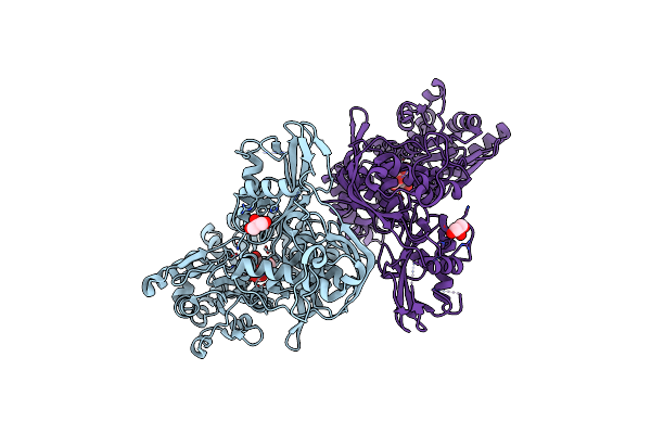
Organism: Geobacillus stearothermophilus, Synthetic construct
Method: X-RAY DIFFRACTION Resolution:3.20 Å Release Date: 2021-08-18 Classification: RNA BINDING PROTEIN/RNA/DNA Ligands: DTP, SO4, MG |
 |
Crystal Structure Of The Cas6 Domain Of Marinomonas Mediterranea Mmb-1 Cas6-Rt-Cas1 Fusion Protein
Organism: Escherichia coli o157:h7, Marinomonas mediterranea mmb-1
Method: X-RAY DIFFRACTION Resolution:2.85 Å Release Date: 2018-10-17 Classification: HYDROLASE Ligands: GOL, SO4 |
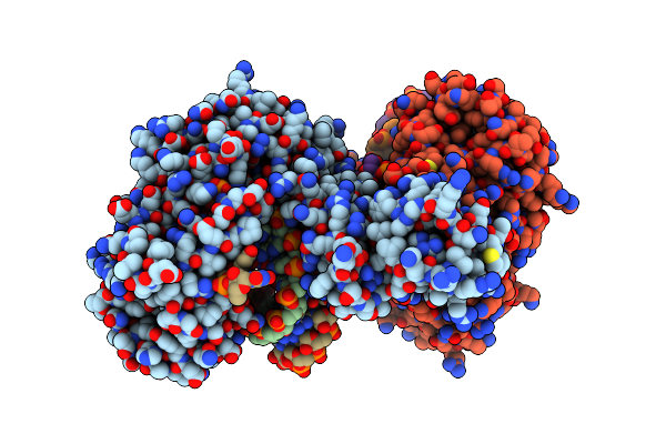 |
Structure Of A Thermostable Group Ii Intron Reverse Transcriptase With Template-Primer And Its Functional And Evolutionary Implications (Rt/Duplex (Nat))
Organism: Geobacillus stearothermophilus, Synthetic construct
Method: X-RAY DIFFRACTION Resolution:3.01 Å Release Date: 2017-11-29 Classification: RNA binding protein/RNA/DNA Ligands: DTP, SO4, MG |
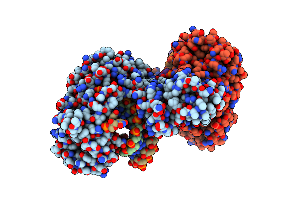 |
Structure Of A Thermostable Group Ii Intron Reverse Transcriptase With Template-Primer And Its Functional And Evolutionary Implications (Rt/Duplex (Se-Met))
Organism: Geobacillus stearothermophilus, Synthetic construct
Method: X-RAY DIFFRACTION Resolution:3.41 Å Release Date: 2017-11-29 Classification: RNA binding protein/RNA/DNA Ligands: DTP, MG |
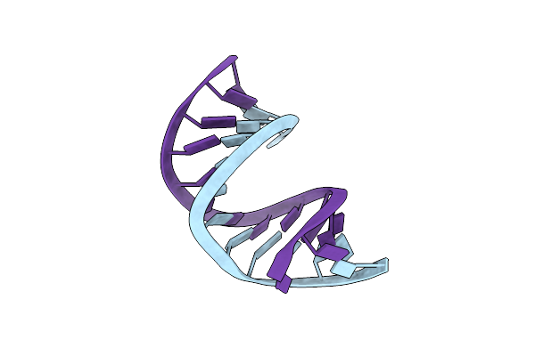 |
Structure Of A Thermostable Group Ii Intron Reverse Transcriptase With Template-Primer And Its Functional And Evolutionary Implications (Duplex Only)
Organism: Synthetic construct
Method: X-RAY DIFFRACTION Resolution:2.41 Å Release Date: 2017-11-29 Classification: DNA/RNA |
 |
Organism: Homo sapiens
Method: X-RAY DIFFRACTION Resolution:2.50 Å Release Date: 2014-03-26 Classification: TRANSFERASE/PEPTIDE Ligands: GOL, MG, ADP, DTT, CL |
 |
Crystal Structure Of Gsk-3/Axin Complex Bound To Phosphorylated N-Terminal Auto-Inhibitory Ps9 Peptide
Organism: Homo sapiens
Method: X-RAY DIFFRACTION Resolution:2.10 Å Release Date: 2014-03-26 Classification: TRANSFERASE/PEPTIDE Ligands: GOL, MG, CL, DTT, ADP |
 |
Crystal Structure Of Gsk-3/Axin Complex Bound To Phosphorylated Wnt Receptor Lrp6 C-Motif
Organism: Homo sapiens
Method: X-RAY DIFFRACTION Resolution:2.30 Å Release Date: 2014-03-26 Classification: TRANSFERASE/PEPTIDE Ligands: ADP, GOL, MG, CL |
 |
Crystal Structure Of Gsk-3/Axin Complex Bound To Phosphorylated Wnt Receptor Lrp6 E-Motif
Organism: Homo sapiens
Method: X-RAY DIFFRACTION Resolution:2.30 Å Release Date: 2014-03-26 Classification: TRANSFERASE/PEPTIDE Ligands: ADP, GOL, MG, CL, DTT |
 |
Crystal Structure Of A Transition State Mimic Of The Gsk-3/Axin Complex Bound To Phosphorylated N-Terminal Auto-Inhibitory Ps9 Peptide
Organism: Mus musculus, Homo sapiens
Method: X-RAY DIFFRACTION Resolution:2.50 Å Release Date: 2014-03-26 Classification: TRANSFERASE/PEPTIDE Ligands: GOL, MG, ADP, AF3, NO3 |
 |
Organism: Homo sapiens
Method: X-RAY DIFFRACTION Resolution:2.70 Å Release Date: 2005-02-15 Classification: HYDROLASE Ligands: NAG |
 |
Organism: Homo sapiens
Method: X-RAY DIFFRACTION Resolution:2.60 Å Release Date: 2005-02-15 Classification: HYDROLASE/INHIBITOR Ligands: PO4 |
 |
The Crystal Structure Of Beta-Catenin Armadillo Repeat Complexed With A Phosphorylated Apc 20Mer Repeat.
Organism: Mus musculus, Homo sapiens
Method: X-RAY DIFFRACTION Resolution:2.10 Å Release Date: 2005-01-12 Classification: SIGNALING PROTEIN |
 |
Crystal Structure Of Beta-Catenin/Icat Helical Domain/Unphosphorylated Apc R3
Organism: Homo sapiens
Method: X-RAY DIFFRACTION Resolution:2.10 Å Release Date: 2004-10-12 Classification: cell adhesion/cell cycle |
 |
High Affinity Ige Receptor (Alpha Chain) Complexed With Tight-Binding E131 'Zeta' Peptide From Phage Display
Organism: Homo sapiens
Method: X-RAY DIFFRACTION Resolution:3.00 Å Release Date: 2004-07-20 Classification: MEMBRANE PROTEIN Ligands: NAG, SO4, CIT |
 |
The Crystal Structure Of Hgf Beta-Chain In Complex With The Sema Domain Of The Met Receptor.
Organism: Homo sapiens
Method: X-RAY DIFFRACTION Resolution:3.22 Å Release Date: 2004-06-15 Classification: GROWTH FACTOR/GROWTH FACTOR RECEPTOR |
 |
Organism: Homo sapiens
Method: X-RAY DIFFRACTION Resolution:2.60 Å Release Date: 2002-09-04 Classification: TRANSFERASE |