
Search Count: 7
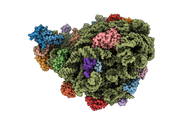 |
Organism: Porphyromonas gingivalis
Method: ELECTRON MICROSCOPY Release Date: 2025-08-20 Classification: RIBOSOME Ligands: MG, ZN, 62B, K, NA |
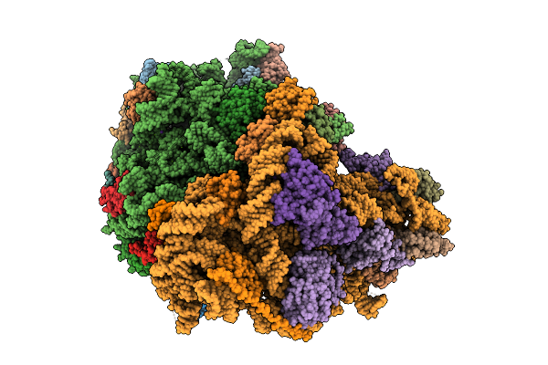 |
Organism: Porphyromonas gingivalis
Method: ELECTRON MICROSCOPY Release Date: 2025-08-20 Classification: RIBOSOME Ligands: MG, K, NA |
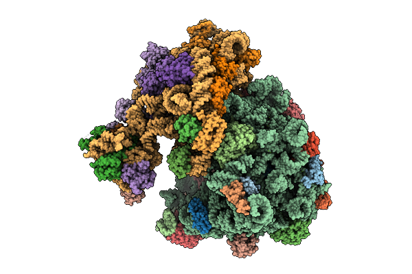 |
Organism: Porphyromonas gingivalis
Method: ELECTRON MICROSCOPY Release Date: 2025-08-20 Classification: RIBOSOME Ligands: MG, ZN, K, NA |
 |
Crystal Structure Of Porphyromonas Gingivalis Peptidylarginine Deiminase (Ppad) Substrate-Unbound.
Organism: Porphyromonas gingivalis w83
Method: X-RAY DIFFRACTION Resolution:1.50 Å Release Date: 2015-07-15 Classification: HYDROLASE |
 |
Crystal Structure Of Porphyromonas Gingivalis Peptidylarginine Deiminase (Ppad) In Complex With Dipeptide Asp-Gln.
Organism: Porphyromonas gingivalis
Method: X-RAY DIFFRACTION Resolution:1.40 Å Release Date: 2015-07-01 Classification: HYDROLASE |
 |
Crystal Structure Of Porphyromonas Gingivalis Peptidylarginine Deiminase (Ppad) Mutant C351A In Complex With Dipeptide Met-Arg.
Organism: Porphyromonas gingivalis (strain atcc baa-308 / w83)
Method: X-RAY DIFFRACTION Resolution:1.80 Å Release Date: 2015-07-01 Classification: HYDROLASE |
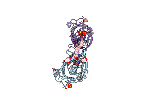 |
Structure Of Porphyromonas Gingivalis Heme-Binding Protein Hmuy In Complex With Heme
Organism: Porphyromonas gingivalis
Method: X-RAY DIFFRACTION Resolution:1.80 Å Release Date: 2009-05-12 Classification: heme-binding protein Ligands: HEM, GOL, SO4 |